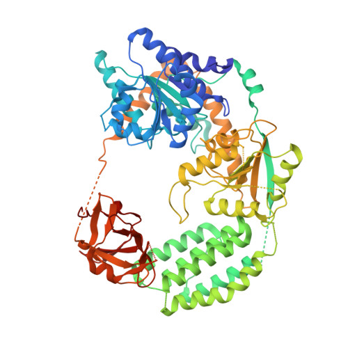
4GL2

Structural Basis for dsRNA duplex backbone recognition by MDA5


Experimental Data Snapshot

  • Method: X-RAY DIFFRACTION
  • Resolution: 3.56 Å
  • R-Value Free: 0.320 
  • R-Value Work: 0.276 
  • R-Value Observed: 0.278 

wwPDB Validation   3D Report Full Report


Ligand Structure Quality Assessment 


This is version 1.2 of the entry. See complete history


Literature

Structural Basis for dsRNA Recognition, Filament Formation, and Antiviral Signal Activation by MDA5.

Wu, B.Peisley, A.Richards, C.Yao, H.Zeng, X.Lin, C.Chu, F.Walz, T.Hur, S.

(2013) Cell 152: 276-289

  • DOI: https://doi.org/10.1016/j.cell.2012.11.048
  • Primary Citation of Related Structures:  
    4GL2

  • PubMed Abstract: 

    MDA5, a viral double-stranded RNA (dsRNA) receptor, shares sequence similarity and signaling pathways with RIG-I yet plays essential functions in antiviral immunity through distinct specificity for viral RNA. Revealing the molecular basis for the functional divergence, we report here the crystal structure of MDA5 bound to dsRNA, which shows how, using the same domain architecture, MDA5 recognizes the internal duplex structure, whereas RIG-I recognizes the terminus of dsRNA. We further show that MDA5 uses direct protein-protein contacts to stack along dsRNA in a head-to-tail arrangement, and that the signaling domain (tandem CARD), which decorates the outside of the core MDA5 filament, also has an intrinsic propensity to oligomerize into an elongated structure that activates the signaling adaptor, MAVS. These data support a model in which MDA5 uses long dsRNA as a signaling platform to cooperatively assemble the core filament, which in turn promotes stochastic assembly of the tandem CARD oligomers for signaling.


  • Organizational Affiliation

    Department of Biological Chemistry and Molecular Pharmacology, Harvard Medical School, Boston, MA 02115, USA.


Macromolecules

Find similar proteins by:  (by identity cutoff)  |  3D Structure
Entity ID: 1
MoleculeChains Sequence LengthOrganismDetailsImage
Interferon-induced helicase C domain-containing protein 1
A, B
699Homo sapiensMutation(s): 0 
Gene Names: IFIH1MDA5RH116
EC: 3.6.4.13
UniProt & NIH Common Fund Data Resources
Find proteins for Q9BYX4 (Homo sapiens)
Explore Q9BYX4 
Go to UniProtKB:  Q9BYX4
PHAROS:  Q9BYX4
GTEx:  ENSG00000115267 
Entity Groups  
Sequence Clusters30% Identity50% Identity70% Identity90% Identity95% Identity100% Identity
UniProt GroupQ9BYX4
Sequence Annotations
Expand
  • Reference Sequence

Find similar nucleic acids by:  Sequence   |   3D Structure  

Entity ID: 2
MoleculeChains LengthOrganismImage
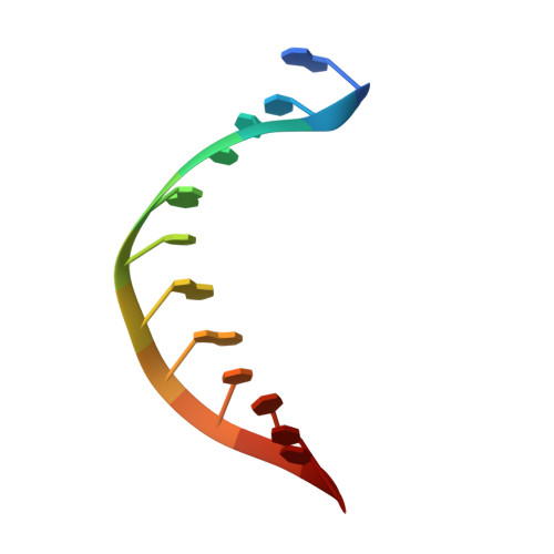
RNA (5'-R(*AP*UP*CP*CP*GP*CP*GP*GP*CP*CP*CP*U)-3')C,
D [auth E]
12N/A
Sequence Annotations
Expand
  • Reference Sequence

Find similar nucleic acids by:  Sequence   |   3D Structure  

Entity ID: 3
MoleculeChains LengthOrganismImage
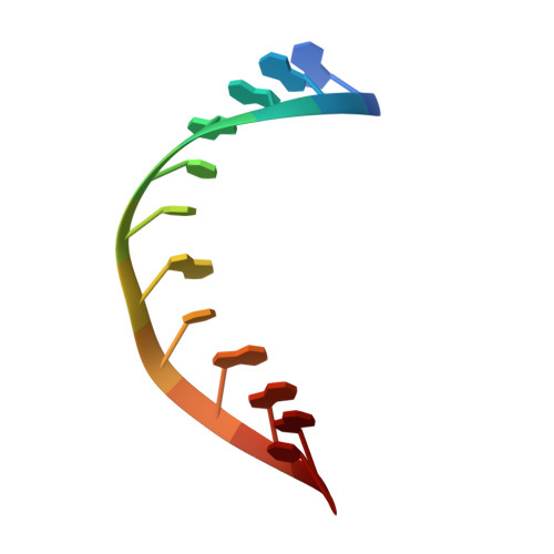
RNA (5'-R(P*AP*GP*GP*GP*CP*CP*GP*CP*GP*GP*AP*U)-3')E [auth F],
F [auth D]
12N/A
Sequence Annotations
Expand
  • Reference Sequence
Experimental Data & Validation

Experimental Data

  • Method: X-RAY DIFFRACTION
  • Resolution: 3.56 Å
  • R-Value Free: 0.320 
  • R-Value Work: 0.276 
  • R-Value Observed: 0.278 
  • Space Group: P 21 21 21
Unit Cell:
Length ( Å )Angle ( ˚ )
a = 71.112α = 90
b = 154.748β = 90
c = 185.006γ = 90
Software Package:
Software NamePurpose
APSdata collection
PHASESphasing
PHENIXrefinement
HKL-2000data reduction
SCALAdata scaling

Structure Validation

View Full Validation Report


Ligand Structure Quality Assessment 


Entry History 

Deposition Data

  • Released Date: 2013-01-09 
  • Deposition Author(s): Wu, B., Hur, S.

Revision History  (Full details and data files)

  • Version 1.0: 2013-01-09
    Type: Initial release
  • Version 1.1: 2013-02-06
    Changes: Database references
  • Version 1.2: 2024-10-30
    Changes: Data collection, Database references, Derived calculations, Structure summary