4GKZ | pdb_00004gkz

HA1.7, a MHC class II restricted TCR specific for haemagglutinin


Experimental Data Snapshot

  • Method: X-RAY DIFFRACTION
  • Resolution: 2.39 Å
  • R-Value Free: 
    0.298 (Depositor), 0.294 (DCC) 
  • R-Value Work: 
    0.249 (Depositor), 0.237 (DCC) 
  • R-Value Observed: 
    0.251 (Depositor) 

Starting Model: experimental
View more details

wwPDB Validation 3D Report Full Report

Validation slider image for 4GKZ

This is version 1.2 of the entry. See complete history

Literature

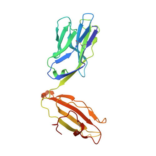
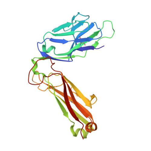
Minimal conformational plasticity enables TCR cross-reactivity to different MHC class II heterodimers.

Holland, C.J.Rizkallah, P.J.Vollers, S.Calvo-Calle, J.M.Madura, F.Fuller, A.Sewell, A.K.Stern, L.J.Godkin, A.Cole, D.K.

(2012) Sci Rep 2: 629-629

  • DOI: https://doi.org/10.1038/srep00629
  • Primary Citation Related Structures: 
    4GKZ

  • PubMed Abstract: 

    Successful immunity requires that a limited pool of αβ T-cell receptors (TCRs) provide cover for a vast number of potential foreign peptide antigens presented by 'self' major histocompatibility complex (pMHC) molecules. Structures of unligated and ligated MHC class-I-restricted TCRs with different ligands, supplemented with biophysical analyses, have revealed a number of important mechanisms that govern TCR mediated antigen recognition. HA1.7 TCR binding to the influenza hemagglutinin antigen (HA(306-318)) presented by HLA-DR1 or HLA-DR4 represents an ideal system for interrogating pMHC-II antigen recognition. Accordingly, we solved the structure of the unligated HA1.7 TCR and compared it to both complex structures. Despite a relatively rigid binding mode, HA1.7 T-cells could tolerate mutations in key contact residues within the peptide epitope. Thermodynamic analysis revealed that limited plasticity and extreme favorable entropy underpinned the ability of the HA1.7 T-cell clone to cross-react with HA(306-318) presented by multiple MHC-II alleles.


  • Organizational Affiliation
    • Institute of Infection and Immunity, Cardiff University School of Medicine, Cardiff, United Kingdom.

Macromolecule Content 

  • Total Structure Weight: 50.63 kDa 
  • Atom Count: 3,626 
  • Modeled Residue Count: 443 
  • Deposited Residue Count: 443 
  • Unique protein chains: 2

Macromolecules

Find similar proteins by:|  3D Structure
Entity ID: 1
MoleculeChains  Sequence LengthOrganismDetailsImage
Alpha chain of Class II TCR202Homo sapiensMutation(s): 0 
Find similar proteins by:|  3D Structure
Entity ID: 2
MoleculeChains  Sequence LengthOrganismDetailsImage
Beta Chain of Class II TCR241Homo sapiensMutation(s): 0 

Small Molecules

Ligands 3 Unique
IDChains Name / Formula / InChI Key2D Diagram3D Interactions
TAM

Query on TAM


Download:Ideal Coordinates CCD File
I [auth A]TRIS(HYDROXYETHYL)AMINOMETHANE
C7 H17 N O3
GKODZWOPPOTFGA-UHFFFAOYSA-N
SO4

Query on SO4


Download:Ideal Coordinates CCD File
G [auth A],
H [auth A]
SULFATE ION
O4 S
QAOWNCQODCNURD-UHFFFAOYSA-L
EDO

Query on EDO


Download:Ideal Coordinates CCD File
C [auth A]
D [auth A]
E [auth A]
F [auth A]
J [auth B]
C [auth A],
D [auth A],
E [auth A],
F [auth A],
J [auth B],
K [auth B]
1,2-ETHANEDIOL
C2 H6 O2
LYCAIKOWRPUZTN-UHFFFAOYSA-N

Experimental Data & Validation

Experimental Data

  • Method: X-RAY DIFFRACTION
  • Resolution: 2.39 Å
  • R-Value Free:  0.298 (Depositor), 0.294 (DCC) 
  • R-Value Work:  0.249 (Depositor), 0.237 (DCC) 
  • R-Value Observed: 0.251 (Depositor) 
Space Group: P 1 21 1
Unit Cell:
Length ( Å )Angle ( ˚ )
a = 69.24α = 90
b = 49.66β = 94.33
c = 72.61γ = 90
Software Package:
Software NamePurpose
SCALAdata scaling
PHASERphasing
REFMACrefinement
PDB_EXTRACTdata extraction
GDAdata collection
xia2data reduction

Structure Validation

View Full Validation Report


Entry History 

Deposition Data

Revision History  (Full details and data files)

  • Version 1.0: 2012-11-07
    Type: Initial release
  • Version 1.1: 2023-09-13
    Changes: Data collection, Database references, Derived calculations, Refinement description
  • Version 1.2: 2024-11-27
    Changes: Structure summary