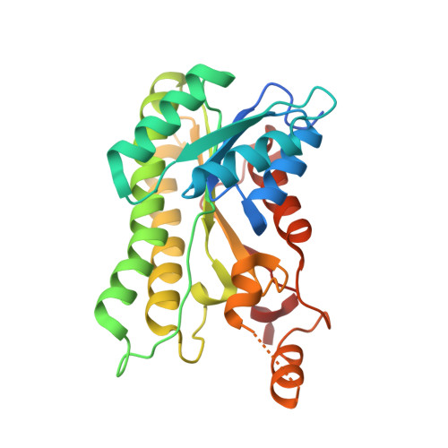
4GH5 | pdb_00004gh5

Crystal structure of S-2-hydroxypropyl coenzyme M dehydrogenase (S-HPCDH)


Experimental Data Snapshot

  • Method: X-RAY DIFFRACTION
  • Resolution: 1.60 Å
  • R-Value Free: 
    0.199 (Depositor), 0.190 (DCC) 
  • R-Value Work: 
    0.171 (Depositor), 0.160 (DCC) 
  • R-Value Observed: 
    0.172 (Depositor) 

Starting Model: experimental
View more details

wwPDB Validation   3D Report Full Report


Ligand Structure Quality Assessment 

Created with Raphaël 2.3.0Worse 01 BetterLigand structure goodness of fit to experimental dataBest fitted NADClick on this verticalbar to view details

This is version 1.2 of the entry. See complete history


Literature

Crystal structures of S-HPCDH reveal determinants of stereospecificity for R- and S-hydroxypropyl-coenzyme M dehydrogenases.

Bakelar, J.W.Sliwa, D.A.Johnson, S.J.

(2013) Arch Biochem Biophys 533: 62-68

  • DOI: https://doi.org/10.1016/j.abb.2013.02.017
  • Primary Citation of Related Structures:  
    4GH5, 4ITU

  • PubMed Abstract: 

    (R)- and (S)-hydroxypropyl-coenzyme M dehydrogenases (R- and S-HPCDH) are stereospecific enzymes that are central to the metabolism of propylene and epoxide in Xanthobacter autotrophicus. The bacterium produces R- and S-HPCDH simultaneously to facilitate transformation of R- and S-enantiomers of epoxypropane to a common achiral product 2-ketopropyl-CoM (2-KPC). Both R- and S-HPCDH are highly specific for their respective substrates as each enzyme displays less than 0.5% activity with the opposite substrate isomer. In order to elucidate the structural basis for stereospecificity displayed by R- and S-HPCDH we have determined substrate bound crystal structures of S-HPCDH to 1.6Å resolution. Comparisons to the previously reported product-bound structure of R-HPCDH reveal that although the placement of catalytic residues within the active site of each enzyme is nearly identical, structural differences in the surrounding area provide each enzyme with a distinct substrate binding pocket. These structures demonstrate how chiral discrimination by R- and S-HPCDH results from alternative binding of the distal end of substrates within each substrate binding pocket.


  • Organizational Affiliation

    Department of Chemistry & Biochemistry, Utah State University, Logan, UT 84322-0300, USA.


Macromolecules
Find similar proteins by:  (by identity cutoff)  |  3D Structure
Entity ID: 1
MoleculeChains Sequence LengthOrganismDetailsImage
Short-chain dehydrogenase/reductase SDR
A, B, C, D
269Xanthobacter autotrophicus Py2Mutation(s): 0 
Gene Names: Xaut_5073
EC: 1.1.1.269
UniProt
Find proteins for A7IQH5 (Xanthobacter autotrophicus (strain ATCC BAA-1158 / Py2))
Explore A7IQH5 
Go to UniProtKB:  A7IQH5
Entity Groups  
Sequence Clusters30% Identity50% Identity70% Identity90% Identity95% Identity100% Identity
UniProt GroupA7IQH5
Sequence Annotations
Expand
  • Reference Sequence
Small Molecules
Ligands 2 Unique
IDChains Name / Formula / InChI Key2D Diagram3D Interactions
NAD
Query on NAD

Download Ideal Coordinates CCD File 
E [auth A],
G [auth B],
I [auth C],
K [auth D]
NICOTINAMIDE-ADENINE-DINUCLEOTIDE
C21 H27 N7 O14 P2
BAWFJGJZGIEFAR-NNYOXOHSSA-N
ACT
Query on ACT

Download Ideal Coordinates CCD File 
F [auth A],
H [auth B],
J [auth C],
L [auth D]
ACETATE ION
C2 H3 O2
QTBSBXVTEAMEQO-UHFFFAOYSA-M
Experimental Data & Validation

Experimental Data

  • Method: X-RAY DIFFRACTION
  • Resolution: 1.60 Å
  • R-Value Free:  0.199 (Depositor), 0.190 (DCC) 
  • R-Value Work:  0.171 (Depositor), 0.160 (DCC) 
  • R-Value Observed: 0.172 (Depositor) 
Space Group: P 21 21 2
Unit Cell:
Length ( Å )Angle ( ˚ )
a = 116.071α = 90
b = 127.921β = 90
c = 58.451γ = 90
Software Package:
Software NamePurpose
CrystalCleardata collection
PHASERphasing
PHENIXrefinement
HKL-2000data reduction
HKL-2000data scaling

Structure Validation

View Full Validation Report


Ligand Structure Quality Assessment 

Created with Raphaël 2.3.0Worse 01 BetterLigand structure goodness of fit to experimental dataBest fitted NADClick on this verticalbar to view details

Entry History 

Deposition Data

Revision History  (Full details and data files)

  • Version 1.0: 2013-03-27
    Type: Initial release
  • Version 1.1: 2013-04-24
    Changes: Database references
  • Version 1.2: 2023-09-13
    Changes: Data collection, Database references, Derived calculations, Refinement description