4GGB | pdb_00004ggb

Crystal structure of a proposed galactarolactone cycloisomerase from agrobacterium tumefaciens, TARGET EFI-500704, WITH BOUND CA, DISORDERED LOOPS


Experimental Data Snapshot

  • Method: X-RAY DIFFRACTION
  • Resolution: 2.00 Å
  • R-Value Free: 
    0.234 (Depositor), 0.233 (DCC) 
  • R-Value Work: 
    0.196 (Depositor), 0.197 (DCC) 
  • R-Value Observed: 
    0.198 (Depositor) 

Starting Model: experimental
View more details

wwPDB Validation 3D Report Full Report

Validation slider image for 4GGB

This is version 1.3 of the entry. See complete history

Literature

Purification, crystallization and structural elucidation of D-galactaro-1,4-lactone cycloisomerase from Agrobacterium tumefaciens involved in pectin degradation.

Vetting, M.W.Bouvier, J.T.Gerlt, J.A.Almo, S.C.

(2016) Acta Crystallogr F Struct Biol Commun 72: 36-41

  • DOI: https://doi.org/10.1107/S2053230X15023286
  • Primary Citation Related Structures: 
    4GGB, 4HPN

  • PubMed Abstract: 

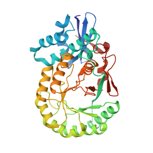
    Pectin is found in the cell wall of plants and is often discarded as waste. A number of research groups are interested in redirecting this biomass waste stream for the production of fuel and bulk chemicals. The primary monomeric subunit of this polysaccharide is D-galacturonate, a six-carbon acid sugar that is degraded in a five-step pathway to central metabolic intermediates by some bacteria, including Agrobacterium tumefaciens. In the third step of the pathway, D-galactaro-1,4-lactone is converted to 2-keto-3-deoxy-L-threo-hexarate by a member of the mandelate racemase subgroup of the enolase superfamily with a novel activity for the superfamily. The 1.6 Å resolution structure of this enzyme was determined, revealing an overall modified (β/α)7β TIM-barrel domain, a hallmark of the superfamily. D-Galactaro-1,4-lactone was manually docked into the active site located at the interface between the N-terminal lid domain and the C-terminal barrel domain. On the basis of the position of the lactone in the active site, Lys166 is predicted to be the active-site base responsible for abstraction of the α proton. His296 on the opposite side of the active site is predicted to be the general acid that donates a proton to the β carbon as the lactone ring opens. The lactone ring appears to be oriented within the active site by stacking interactions with Trp298.


  • Organizational Affiliation
    • Department of Biochemistry, Albert Einstein College of Medicine, Bronx, NY 10461, USA.

Macromolecule Content 

  • Total Structure Weight: 41.8 kDa 
  • Atom Count: 2,869 
  • Modeled Residue Count: 348 
  • Deposited Residue Count: 378 
  • Unique protein chains: 1

Macromolecules

Find similar proteins by:|  3D Structure
Entity ID: 1
MoleculeChains  Sequence LengthOrganismDetailsImage
Putative uncharacterized protein378Agrobacterium fabrum str. C58Mutation(s): 0 
Gene Names: Atu3139
EC: 5.5.1.27
UniProt
Find proteins for A9CEQ8 (Agrobacterium fabrum (strain C58 / ATCC 33970))
Explore A9CEQ8 
Go to UniProtKB:  A9CEQ8
Entity Groups
Sequence Clusters30% Identity50% Identity70% Identity90% Identity95% Identity100% Identity
UniProt GroupA9CEQ8
Sequence Annotations
Expand
Reference Sequence

Small Molecules

Ligands 2 Unique
IDChains Name / Formula / InChI Key2D Diagram3D Interactions
CA

Query on CA


Download:Ideal Coordinates CCD File
B [auth A],
C [auth A],
D [auth A]
CALCIUM ION
Ca
BHPQYMZQTOCNFJ-UHFFFAOYSA-N
CL

Query on CL


Download:Ideal Coordinates CCD File
E [auth A],
F [auth A],
G [auth A],
H [auth A],
I [auth A]
CHLORIDE ION
Cl
VEXZGXHMUGYJMC-UHFFFAOYSA-M

Experimental Data & Validation

Experimental Data

  • Method: X-RAY DIFFRACTION
  • Resolution: 2.00 Å
  • R-Value Free:  0.234 (Depositor), 0.233 (DCC) 
  • R-Value Work:  0.196 (Depositor), 0.197 (DCC) 
  • R-Value Observed: 0.198 (Depositor) 
Space Group: I 4 2 2
Unit Cell:
Length ( Å )Angle ( ˚ )
a = 132.178α = 90
b = 132.178β = 90
c = 95.472γ = 90
Software Package:
Software NamePurpose
SCALEPACKdata scaling
PHENIXrefinement
PDB_EXTRACTdata extraction
MOSFLMdata reduction
PHENIXphasing

Structure Validation

View Full Validation Report


Entry History 

Revision History  (Full details and data files)

  • Version 1.0: 2012-08-29
    Type: Initial release
  • Version 1.1: 2016-06-22
    Changes: Database references
  • Version 1.2: 2016-07-27
    Changes: Database references
  • Version 1.3: 2023-09-13
    Changes: Data collection, Database references, Derived calculations, Refinement description