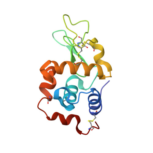
The crystal structure analysis of the relative binding of cisplatin and carboplatin in a mixture with histidine in a protein studied at 100 and 300 K with repeated X-ray irradiation.
Helliwell, J.R., Tanley, S.W.(2013) Acta Crystallogr D Biol Crystallogr 69: 121-125
- PubMed: 23275170
- DOI: https://doi.org/10.1107/S090744491204423X
- Primary Citation of Related Structures:
4GCB, 4GCC, 4GCD, 4GCE, 4GCF - PubMed Abstract:
The anticancer agents cisplatin and carboplatin bind to histidine in a protein. This crystal structure study at data-collection temperatures of 100 and 300 K examines their relative binding affinities to a histidine side chain and the effect of a high X-ray radiation dose of up to ∼1.8 MGy on the stability of the subsequent protein-Pt adducts. Cisplatin binding is visible at the histidine residue, but carboplatin binding is not. Five refined X-ray crystal structures are presented: one at 100 K as a reference and four at 300 K. The diffraction resolutions are 1.8, 2.0, 2.8, 2.9 and 3.5 Å.
- School of Chemistry, University of Manchester, England, UK. john.helliwell@manchester.ac.uk
Organizational Affiliation:


