4GBM | pdb_00004gbm

Sulfotransferase Domain from the Curacin Biosynthetic Pathway


Experimental Data Snapshot

  • Method: X-RAY DIFFRACTION
  • Resolution: 1.62 Å
  • R-Value Free: 
    0.182 (Depositor), 0.181 (DCC) 
  • R-Value Work: 
    0.158 (Depositor), 0.157 (DCC) 
  • R-Value Observed: 
    0.160 (Depositor) 

wwPDB Validation 3D Report Full Report

Validation slider image for 4GBM

Ligand Structure Quality Assessment 


This is version 1.2 of the entry. See complete history

Literature

Structural basis of functional group activation by sulfotransferases in complex metabolic pathways.

McCarthy, J.G.Eisman, E.B.Kulkarni, S.Gerwick, L.Gerwick, W.H.Wipf, P.Sherman, D.H.Smith, J.L.

(2012) ACS Chem Biol 7: 1994-2003

  • DOI: https://doi.org/10.1021/cb300385m
  • Primary Citation Related Structures: 
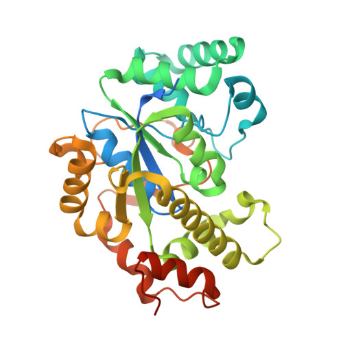
    4GBM, 4GOX

  • PubMed Abstract: 

    Sulfated molecules with diverse functions are common in biology, but sulfonation as a method to activate a metabolite for chemical catalysis is rare. Catalytic activity was characterized and crystal structures were determined for two such "activating" sulfotransferases (STs) that sulfonate β-hydroxyacyl thioester substrates. The CurM polyketide synthase (PKS) ST domain from the curacin A biosynthetic pathway of Moorea producens and the olefin synthase (OLS) ST from a hydrocarbon-producing system of Synechococcus PCC 7002 both occur as a unique acyl carrier protein (ACP), ST, and thioesterase (TE) tridomain within a larger polypeptide. During pathway termination, these cyanobacterial systems introduce a terminal double bond into the β-hydroxyacyl-ACP-linked substrate by the combined action of the ST and TE. Under in vitro conditions, CurM PKS ST and OLS ST acted on β-hydroxy fatty acyl-ACP substrates; however, OLS ST was not reactive toward analogues of the natural PKS ST substrate bearing a C5-methoxy substituent. The crystal structures of CurM ST and OLS ST revealed that they are members of a distinct protein family relative to other prokaryotic and eukaryotic sulfotransferases. A common binding site for the sulfonate donor 3'-phosphoadenosine-5'-phosphosulfate was visualized in complexes with the product 3'-phosphoadenosine-5'-phosphate. Critical functions for several conserved amino acids in the active site were confirmed by site-directed mutagenesis, including a proposed glutamate catalytic base. A dynamic active-site flap unique to the "activating" ST family affects substrate selectivity and product formation, based on the activities of chimeras of the PKS and OLS STs with exchanged active-site flaps.


  • Organizational Affiliation
    • Life Sciences Institute and Department of Biological Chemistry, University of Michigan, Ann Arbor, MI 48109, USA.

Macromolecule Content 

  • Total Structure Weight: 38.14 kDa 
  • Atom Count: 2,773 
  • Modeled Residue Count: 283 
  • Deposited Residue Count: 323 
  • Unique protein chains: 1

Macromolecules

Find similar proteins by:|  3D Structure
Entity ID: 1
MoleculeChains  Sequence LengthOrganismDetailsImage
CurM Sulfotransferase323Moorena producensMutation(s): 2 
Gene Names: CurM
UniProt
Find proteins for F4Y423 (Moorena producens 3L)
Explore F4Y423 
Go to UniProtKB:  F4Y423
Entity Groups
Sequence Clusters30% Identity50% Identity70% Identity90% Identity95% Identity100% Identity
UniProt GroupF4Y423
Sequence Annotations
Expand
Reference Sequence

Small Molecules

Ligands 5 Unique
IDChains Name / Formula / InChI Key2D Diagram3D Interactions
A3P

Query on A3P


Download:Ideal Coordinates CCD File
G [auth A]ADENOSINE-3'-5'-DIPHOSPHATE
C10 H15 N5 O10 P2
WHTCPDAXWFLDIH-KQYNXXCUSA-N
P6G

Query on P6G


Download:Ideal Coordinates CCD File
R [auth A],
S [auth A]
HEXAETHYLENE GLYCOL
C12 H26 O7
IIRDTKBZINWQAW-UHFFFAOYSA-N
SO4

Query on SO4


Download:Ideal Coordinates CCD File
Q [auth A]SULFATE ION
O4 S
QAOWNCQODCNURD-UHFFFAOYSA-L
ZN

Query on ZN


Download:Ideal Coordinates CCD File
B [auth A],
C [auth A],
D [auth A],
E [auth A],
F [auth A]
ZINC ION
Zn
PTFCDOFLOPIGGS-UHFFFAOYSA-N
CL

Query on CL


Download:Ideal Coordinates CCD File
H [auth A]
I [auth A]
J [auth A]
K [auth A]
L [auth A]
H [auth A],
I [auth A],
J [auth A],
K [auth A],
L [auth A],
M [auth A],
N [auth A],
O [auth A],
P [auth A]
CHLORIDE ION
Cl
VEXZGXHMUGYJMC-UHFFFAOYSA-M

Experimental Data & Validation

Experimental Data

  • Method: X-RAY DIFFRACTION
  • Resolution: 1.62 Å
  • R-Value Free:  0.182 (Depositor), 0.181 (DCC) 
  • R-Value Work:  0.158 (Depositor), 0.157 (DCC) 
  • R-Value Observed: 0.160 (Depositor) 
Space Group: P 21 21 21
Unit Cell:
Length ( Å )Angle ( ˚ )
a = 45.856α = 90
b = 67.299β = 90
c = 118.016γ = 90
Software Package:
Software NamePurpose
DENZOdata reduction
SCALEPACKdata scaling
PHASERphasing
RESOLVEphasing
REFMACrefinement
PDB_EXTRACTdata extraction
HKL-2000data collection
HKL-2000data reduction
HKL-2000data scaling

Structure Validation

View Full Validation Report


Ligand Structure Quality Assessment 


Entry History 

Deposition Data

Revision History  (Full details and data files)

  • Version 1.0: 2012-10-17
    Type: Initial release
  • Version 1.1: 2013-01-02
    Changes: Database references
  • Version 1.2: 2024-02-28
    Changes: Data collection, Database references, Derived calculations