4GBF | pdb_00004gbf

Crystal structure of the C-terminal domain of gp131 from bacteriophage phiKZ


Experimental Data Snapshot

  • Method: X-RAY DIFFRACTION
  • Resolution: 1.95 Å
  • R-Value Free: 
    0.199 (Depositor), 0.196 (DCC) 
  • R-Value Work: 
    0.161 (Depositor), 0.158 (DCC) 
  • R-Value Observed: 
    0.163 (Depositor) 

wwPDB Validation 3D Report Full Report

Validation slider image for 4GBF

This is version 1.3 of the entry. See complete history

Literature

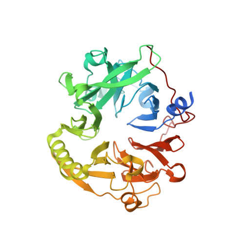
Crystal structure and location of gp131 in the bacteriophage phiKZ virion.

Sycheva, L.V.Shneider, M.M.Sykilinda, N.N.Ivanova, M.A.Miroshnikov, K.A.Leiman, P.G.

(2012) Virology 434: 257-264

  • DOI: https://doi.org/10.1016/j.virol.2012.09.001
  • Primary Citation Related Structures: 
    4GBF

  • PubMed Abstract: 

    Pseudomonas phage ϕKZ and its two close relatives ϕPA3 and 201ϕ2-1 are very large bacteriophages that form a separate branch in phage classification because their genomes are very different from the rest of GenBank sequence data. The contractile tail of ϕKZ is built from at least 32 different proteins, but a definitive structural function is assigned to only one of them-the tail sheath protein. Here, we report the crystal structure of the C-terminal domain of another phiKZ tail protein, gene product 131 (gp131C). We show that gp131 is located at the periphery of the baseplate and possibly associates with fibers that emanate from the baseplate. Gp131C is a seven-bladed β-propeller that has a shape of a skewed toroid. A small but highly conserved and negatively charged patch on the surface of gp131C might be important for substrate binding or for interaction with a different tail protein.


  • Organizational Affiliation
    • École Polytechnique Fédérale de Lausanne, Laboratory of Structural Biology and Biophysics, BSP-415, 1015 Lausanne, Switzerland.

Macromolecule Content 

  • Total Structure Weight: 88.19 kDa 
  • Atom Count: 6,447 
  • Modeled Residue Count: 739 
  • Deposited Residue Count: 800 
  • Unique protein chains: 1

Macromolecules

Find similar proteins by:|  3D Structure
Entity ID: 1
MoleculeChains  Sequence LengthOrganismDetailsImage
PHIKZ131
A, B
400Phikzvirus phiKZMutation(s): 0 
Gene Names: ORF131
UniProt
Find proteins for Q8SD31 (Pseudomonas phage phiKZ)
Explore Q8SD31 
Go to UniProtKB:  Q8SD31
Entity Groups
Sequence Clusters30% Identity50% Identity70% Identity90% Identity95% Identity100% Identity
UniProt GroupQ8SD31
Sequence Annotations
Expand
Reference Sequence

Small Molecules

Ligands 2 Unique
IDChains Name / Formula / InChI Key2D Diagram3D Interactions
CL

Query on CL


Download:Ideal Coordinates CCD File
C [auth A],
D [auth A],
H [auth B],
I [auth B]
CHLORIDE ION
Cl
VEXZGXHMUGYJMC-UHFFFAOYSA-M
MG

Query on MG


Download:Ideal Coordinates CCD File
E [auth A],
F [auth A],
G [auth A],
J [auth B],
K [auth B]
MAGNESIUM ION
Mg
JLVVSXFLKOJNIY-UHFFFAOYSA-N

Experimental Data & Validation

Experimental Data

  • Method: X-RAY DIFFRACTION
  • Resolution: 1.95 Å
  • R-Value Free:  0.199 (Depositor), 0.196 (DCC) 
  • R-Value Work:  0.161 (Depositor), 0.158 (DCC) 
  • R-Value Observed: 0.163 (Depositor) 
Space Group: P 21 21 21
Unit Cell:
Length ( Å )Angle ( ˚ )
a = 79.449α = 90
b = 86.353β = 90
c = 97.362γ = 90
Software Package:
Software NamePurpose
RemDAqdata collection
SHELXmodel building
PHENIXrefinement
XDSdata reduction
SCALAdata scaling
SHELXphasing

Structure Validation

View Full Validation Report


Entry History 

Deposition Data

Revision History  (Full details and data files)

  • Version 1.0: 2012-10-17
    Type: Initial release
  • Version 1.1: 2013-01-02
    Changes: Database references
  • Version 1.2: 2017-11-15
    Changes: Refinement description
  • Version 1.3: 2024-02-28
    Changes: Data collection, Database references, Derived calculations