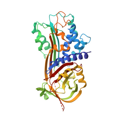
4G8O

Crystal Structure of a novel small molecule inactivator bound to plasminogen activator inhibitor-1


Experimental Data Snapshot

  • Method: X-RAY DIFFRACTION
  • Resolution: 2.71 Å
  • R-Value Free: 0.268 
  • R-Value Work: 0.239 
  • R-Value Observed: 0.241 

wwPDB Validation   3D Report Full Report


Ligand Structure Quality Assessment 


This is version 1.3 of the entry. See complete history


Literature

Mechanistic characterization and crystal structure of a small molecule inactivator bound to plasminogen activator inhibitor-1.

Li, S.H.Reinke, A.A.Sanders, K.L.Emal, C.D.Whisstock, J.C.Stuckey, J.A.Lawrence, D.A.

(2013) Proc Natl Acad Sci U S A 110: E4941-E4949

  • DOI: https://doi.org/10.1073/pnas.1216499110
  • Primary Citation of Related Structures:  
    4G8O, 4G8R

  • PubMed Abstract: 

    Plasminogen activator inhibitor type-1 (PAI-1) is a member of the serine protease inhibitor (serpin) family. Excessive PAI-1 activity is associated with human disease, making it an attractive pharmaceutical target. However, like other serpins, PAI-1 has a labile structure, making it a difficult target for the development of small molecule inhibitors, and to date, there are no US Food and Drug Administration-approved small molecule inactivators of any serpins. Here we describe the mechanistic and structural characterization of a high affinity inactivator of PAI-1. This molecule binds to PAI-1 reversibly and acts through an allosteric mechanism that inhibits PAI-1 binding to proteases and to its cofactor vitronectin. The binding site is identified by X-ray crystallography and mutagenesis as a pocket at the interface of β-sheets B and C and α-helix H. A similar pocket is present on other serpins, suggesting that this site could be a common target in this structurally conserved protein family.


  • Organizational Affiliation

    Departments of Pathology and Internal Medicine, Division of Cardiovascular Medicine, University of Michigan Medical School, Ann Arbor, MI 48109.


Macromolecules
Find similar proteins by:  (by identity cutoff)  |  3D Structure
Entity ID: 1
MoleculeChains Sequence LengthOrganismDetailsImage
Plasminogen activator inhibitor 1
A, B, C, D
376Homo sapiensMutation(s): 4 
Gene Names: PAI-1PAI1PLANH1SERPINE1
UniProt & NIH Common Fund Data Resources
Find proteins for P05121 (Homo sapiens)
Explore P05121 
Go to UniProtKB:  P05121
PHAROS:  P05121
GTEx:  ENSG00000106366 
Entity Groups  
Sequence Clusters30% Identity50% Identity70% Identity90% Identity95% Identity100% Identity
UniProt GroupP05121
Sequence Annotations
Expand
  • Reference Sequence
Small Molecules
Ligands 3 Unique
IDChains Name / Formula / InChI Key2D Diagram3D Interactions
96P
Query on 96P

Download Ideal Coordinates CCD File 
L [auth D](2S)-3-({[3-(trifluoromethyl)phenoxy]carbonyl}amino)propane-1,2-diyl bis(3,4,5-trihydroxybenzoate)
C25 H20 F3 N O12
OXWKLJPAKUDPJY-HNNXBMFYSA-N
SO4
Query on SO4

Download Ideal Coordinates CCD File 
E [auth A]
G [auth B]
H [auth C]
I [auth C]
J [auth D]
E [auth A],
G [auth B],
H [auth C],
I [auth C],
J [auth D],
K [auth D]
SULFATE ION
O4 S
QAOWNCQODCNURD-UHFFFAOYSA-L
EDO
Query on EDO

Download Ideal Coordinates CCD File 
F [auth A]1,2-ETHANEDIOL
C2 H6 O2
LYCAIKOWRPUZTN-UHFFFAOYSA-N
Binding Affinity Annotations 
IDSourceBinding Affinity
96P PDBBind:  4G8O Kd: 22 (nM) from 1 assay(s)
Experimental Data & Validation

Experimental Data

  • Method: X-RAY DIFFRACTION
  • Resolution: 2.71 Å
  • R-Value Free: 0.268 
  • R-Value Work: 0.239 
  • R-Value Observed: 0.241 
  • Space Group: P 1
Unit Cell:
Length ( Å )Angle ( ˚ )
a = 65.335α = 91.32
b = 75.317β = 93.49
c = 105.002γ = 115.65
Software Package:
Software NamePurpose
DENZOdata reduction
SCALEPACKdata scaling
BUSTER-TNTrefinement
PDB_EXTRACTdata extraction
MD2data collection
HKL-2000data reduction
HKL-2000data scaling
BUSTERrefinement

Structure Validation

View Full Validation Report


Ligand Structure Quality Assessment 


Entry History 

Deposition Data

Revision History  (Full details and data files)

  • Version 1.0: 2013-12-25
    Type: Initial release
  • Version 1.1: 2014-01-01
    Changes: Database references
  • Version 1.2: 2017-11-15
    Changes: Advisory, Refinement description
  • Version 1.3: 2024-02-28
    Changes: Advisory, Data collection, Database references, Derived calculations