


Enantiomer-dependent amino acid binding affinity of OmpA-like domains from Acinetobacter baumannii peptidoglycan-associated lipoprotein and OmpA
Lee, W.C., Park, J.S., Song, J.H., Kim, S.I., Lee, J.C., Cheong, J., Kim, H.Y.To be published.
Experimental Data Snapshot
Entity ID: 1 | |||||
|---|---|---|---|---|---|
| Molecule | Chains | Sequence Length | Organism | Details | Image |
| Outer membrane protein Omp38 | 123 | Acinetobacter baumannii | Mutation(s): 0 Gene Names: omp38, ompA | 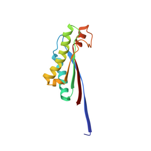 | |
UniProt | |||||
Entity Groups | |||||
| Sequence Clusters | 30% Identity50% Identity70% Identity90% Identity95% Identity100% Identity | ||||
| UniProt Group | Q6RYW5 | ||||
Sequence AnnotationsExpand | |||||
Reference Sequence | |||||
| Ligands 2 Unique | |||||
|---|---|---|---|---|---|
| ID | Chains | Name / Formula / InChI Key | 2D Diagram | 3D Interactions | |
| API Download:Ideal Coordinates CCD File | I [auth A] J [auth B] K [auth C] L [auth D] N [auth E] | 2,6-DIAMINOPIMELIC ACID C7 H14 N2 O4 GMKMEZVLHJARHF-SYDPRGILSA-N |  | ||
| SRT Download:Ideal Coordinates CCD File | M [auth D], Q [auth G] | S,R MESO-TARTARIC ACID C4 H6 O6 FEWJPZIEWOKRBE-XIXRPRMCSA-N |  | ||
| Length ( Å ) | Angle ( ˚ ) |
|---|---|
| a = 58.52 | α = 90 |
| b = 99.3 | β = 105.95 |
| c = 98.12 | γ = 90 |
| Software Name | Purpose |
|---|---|
| SCALEPACK | data scaling |
| MOLREP | phasing |
| CNS | refinement |
| PDB_EXTRACT | data extraction |
| SERGUI | data collection |


