4G6A | pdb_00004g6a

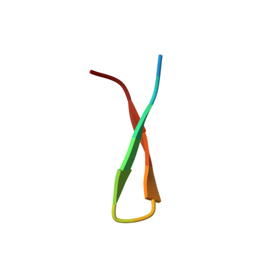
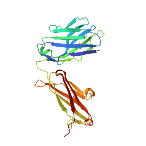
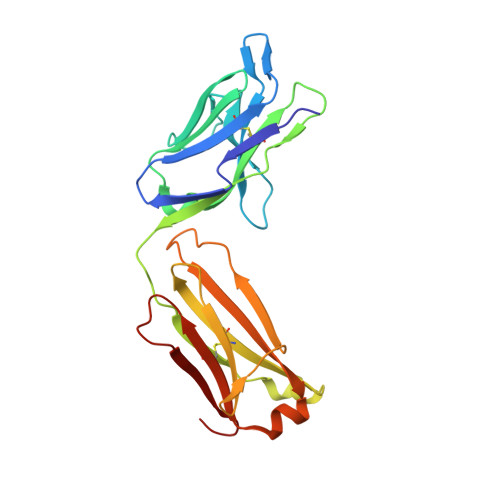
Structure of the Hepatitis C virus envelope glycoprotein E2 antigenic region 412-423 bound to the broadly neutralizing antibody AP33


Experimental Data Snapshot

  • Method: X-RAY DIFFRACTION
  • Resolution: 2.50 Å
  • R-Value Free: 
    0.220 (Depositor), 0.211 (DCC) 
  • R-Value Work: 
    0.178 (Depositor), 0.169 (DCC) 
  • R-Value Observed: 
    0.180 (Depositor) 

Starting Model: experimental
View more details

wwPDB Validation 3D Report Full Report

Validation slider image for 4G6A

This is version 1.3 of the entry. See complete history. 

Literature

Structure of Hepatitis C Virus Envelope Glycoprotein E2 Antigenic Site 412 to 423 in Complex with Antibody AP33.

Kong, L., Giang, E., Nieusma, T., Robbins, J.B., Deller, M.C., Stanfield, R.L., Wilson, I.A., Law, M.

(2012) J Virol 86: 13085-13088

  • DOI: https://doi.org/10.1128/JVI.01939-12
  • Primary Citation Related Structures: 
    4G6A

  • PubMed Abstract: 

    We have determined the crystal structure of the broadly neutralizing antibody (bnAb) AP33, bound to a peptide corresponding to hepatitis C virus (HCV) E2 envelope glycoprotein antigenic site 412 to 423. Comparison with bnAb HCV1 bound to the same epitope reveals a different angle of approach to the antigen by bnAb AP33 and slight variation in its β-hairpin conformation of the epitope. These structures establish two different modes of binding to E2 that antibodies adopt to neutralize diverse HCV.


  • Organizational Affiliation: 
    • Department of Molecular Biology, The Scripps Research Institute, La Jolla, California, USA.

Macromolecule Content 

  • Total Structure Weight: 98.74 kDa 
  • Atom Count: 7,073 
  • Modeled Residue Count: 901 
  • Deposited Residue Count: 910 
  • Unique protein chains: 3

Macromolecules

Find similar proteins by:  Sequence   |   3D Structure  
Entity ID: 1
MoleculeChains  Sequence LengthOrganismDetailsImage
E2 peptideA [auth B],
B [auth A]
13Orthohepacivirus hominisMutation(s): 0 
UniProt
Find proteins for Q9YK84 (Orthohepacivirus hominis)
Explore Q9YK84 
Go to UniProtKB:  Q9YK84
Entity Groups
Sequence Clusters30% Identity50% Identity70% Identity90% Identity95% Identity100% Identity
UniProt GroupQ9YK84
Sequence Annotations
Expand
Reference Sequence
Find similar proteins by:|  3D Structure
Entity ID: 2
MoleculeChains  Sequence LengthOrganismDetailsImage
AP33 Heavy ChainC,
E [auth H]
224Homo sapiensMutation(s): 0 
Entity Groups
Sequence Clusters30% Identity50% Identity70% Identity90% Identity95% Identity100% Identity
Sequence Annotations
Expand
Reference Sequence
Find similar proteins by:|  3D Structure
Entity ID: 3
MoleculeChains  Sequence LengthOrganismDetailsImage
AP33 Light chainD,
F [auth L]
218Homo sapiensMutation(s): 0 
Entity Groups
Sequence Clusters30% Identity50% Identity70% Identity90% Identity95% Identity100% Identity
Sequence Annotations
Expand
Reference Sequence

Experimental Data & Validation

Experimental Data

  • Method: X-RAY DIFFRACTION
  • Resolution: 2.50 Å
  • R-Value Free:  0.220 (Depositor), 0.211 (DCC) 
  • R-Value Work:  0.178 (Depositor), 0.169 (DCC) 
  • R-Value Observed: 0.180 (Depositor) 
Space Group: P 1 21 1
Unit Cell:
Length ( Å )Angle ( ˚ )
a = 44.875α = 90
b = 191.804β = 111.08
c = 59.374γ = 90
Software Package:
Software NamePurpose
Blu-Icedata collection
PHASERphasing
PHENIXrefinement
HKL-2000data reduction
SCALEPACKdata scaling

Structure Validation

View Full Validation Report


Entry History 

Deposition Data

Revision History  (Full details and data files)

  • Version 1.0: 2012-09-26
    Type: Initial release
  • Version 1.1: 2012-11-14
    Changes: Database references
  • Version 1.2: 2023-09-13
    Changes: Data collection, Database references, Refinement description
  • Version 1.3: 2024-10-30
    Changes: Structure summary