4FXL | pdb_00004fxl

Crystal structure of the D76N Beta-2 Microglobulin mutant


Experimental Data Snapshot

  • Method: X-RAY DIFFRACTION
  • Resolution: 1.40 Å
  • R-Value Free: 
    0.186 (Depositor), 0.192 (DCC) 
  • R-Value Work: 
    0.130 (Depositor), 0.139 (DCC) 
  • R-Value Observed: 
    0.133 (Depositor) 

Starting Model: experimental
View more details

wwPDB Validation 3D Report Full Report

Validation slider image for 4FXL

This is version 1.2 of the entry. See complete history

Literature

Hereditary systemic amyloidosis due to Asp76Asn variant beta-2-microglobulin.

Valleix, S.Gillmore, J.D.Bridoux, F.Mangione, P.P.Dogan, A.Nedelec, B.Boimard, M.Touchard, G.Goujon, J.M.Lacombe, C.Lozeron, P.Adams, D.Lacroix, C.Maisonobe, T.Plante-Bordeneuve, V.Vrana, J.A.Theis, J.D.Giorgetti, S.Porcari, R.Ricagno, S.Bolognesi, M.Stoppini, M.Delpech, M.Pepys, M.B.Hawkins, P.N.Bellotti, V.

(2012) N Engl J Med 366: 2276-2283

  • DOI: https://doi.org/10.1056/NEJMoa1201356
  • Primary Citation Related Structures: 
    4FXL

  • PubMed Abstract: 

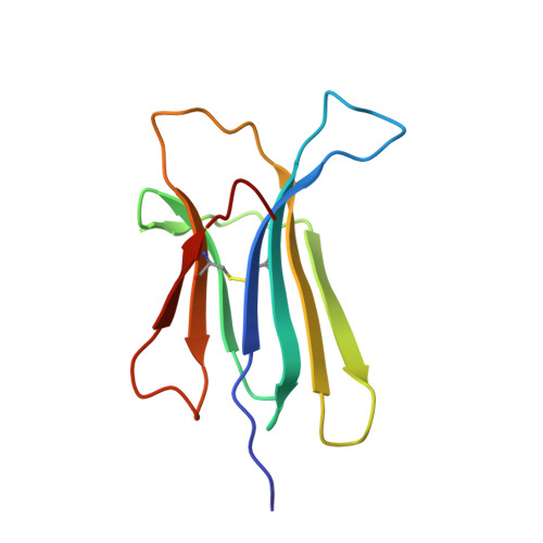
    We describe a kindred with slowly progressive gastrointestinal symptoms and autonomic neuropathy caused by autosomal dominant, hereditary systemic amyloidosis. The amyloid consists of Asp76Asn variant β(2)-microglobulin. Unlike patients with dialysis-related amyloidosis caused by sustained high plasma concentrations of wild-type β(2)-microglobulin, the affected members of this kindred had normal renal function and normal circulating β(2)-microglobulin values. The Asp76Asn β(2)-microglobulin variant was thermodynamically unstable and remarkably fibrillogenic in vitro under physiological conditions. Previous studies of β(2)-microglobulin aggregation have not shown such amyloidogenicity for single-residue substitutions. Comprehensive biophysical characterization of the β(2)-microglobulin variant, including its 1.40-Å, three-dimensional structure, should allow further elucidation of fibrillogenesis and protein misfolding.


  • Organizational Affiliation
    • Laboratoire de Biochimie et de Génétique Moléculaire, Université Paris-Descartes, Sorbonne Paris Cité, Faculté de Médecine Paris, Assistance Public–Hôpitaux de Paris (AP-HP), Paris, France. sophie.valleix@cch.aphp.fr

Macromolecule Content 

  • Total Structure Weight: 12.07 kDa 
  • Atom Count: 982 
  • Modeled Residue Count: 100 
  • Deposited Residue Count: 100 
  • Unique protein chains: 1

Macromolecules

Find similar proteins by:|  3D Structure
Entity ID: 1
MoleculeChains  Sequence LengthOrganismDetailsImage
Beta-2-microglobulin100Homo sapiensMutation(s): 1 
Gene Names: B2MCDABP0092HDCMA22P
UniProt & NIH Common Fund Data Resources
Find proteins for P61769 (Homo sapiens)
Explore P61769 
Go to UniProtKB:  P61769
PHAROS:  P61769
GTEx:  ENSG00000166710 
Entity Groups
Sequence Clusters30% Identity50% Identity70% Identity90% Identity95% Identity100% Identity
UniProt GroupP61769
Sequence Annotations
Expand
Reference Sequence

Experimental Data & Validation

Experimental Data

  • Method: X-RAY DIFFRACTION
  • Resolution: 1.40 Å
  • R-Value Free:  0.186 (Depositor), 0.192 (DCC) 
  • R-Value Work:  0.130 (Depositor), 0.139 (DCC) 
  • R-Value Observed: 0.133 (Depositor) 
Space Group: C 1 2 1
Unit Cell:
Length ( Å )Angle ( ˚ )
a = 76.22α = 90
b = 28.42β = 126.54
c = 52.26γ = 90
Software Package:
Software NamePurpose
MxCuBEdata collection
PHASERphasing
REFMACrefinement
MOSFLMdata reduction
SCALAdata scaling

Structure Validation

View Full Validation Report


Entry History 

Deposition Data

Revision History  (Full details and data files)

  • Version 1.0: 2012-08-15
    Type: Initial release
  • Version 1.1: 2023-09-13
    Changes: Data collection, Database references, Derived calculations, Refinement description
  • Version 1.2: 2024-11-27
    Changes: Structure summary