4FWG | pdb_00004fwg

Crystal structure of the Lon-like protease MtaLonC in complex with lactacystin


Experimental Data Snapshot

  • Method: X-RAY DIFFRACTION
  • Resolution: 1.99 Å
  • R-Value Free: 
    0.259 (Depositor), 0.257 (DCC) 
  • R-Value Work: 
    0.230 (Depositor), 0.218 (DCC) 
  • R-Value Observed: 
    0.231 (Depositor) 

Starting Model: experimental
View more details

wwPDB Validation 3D Report Full Report

Validation slider image for 4FWG

Ligand Structure Quality Assessment 


This is version 1.5 of the entry. See complete history

Literature

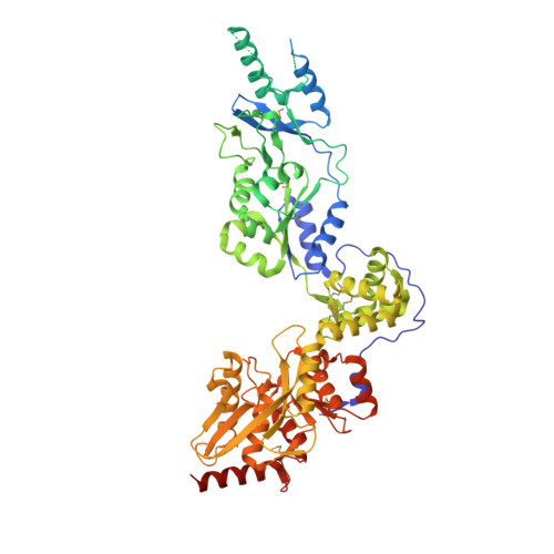
Structures of an ATP-independent Lon-like protease and its complexes with covalent inhibitors

Liao, J.H.Ihara, K.Kuo, C.I.Huang, K.F.Wakatsuki, S.Wu, S.H.Chang, C.I.

(2013) Acta Crystallogr D Biol Crystallogr 69: 1395-1402

  • DOI: https://doi.org/10.1107/S0907444913008214
  • Primary Citation Related Structures: 
    4FW9, 4FWD, 4FWG, 4FWH

  • PubMed Abstract: 

    The Lon proteases are a unique family of chambered proteases with a built-in AAA+ (ATPases associated with diverse cellular activities) module. Here, crystal structures of a unique member of the Lon family with no intrinsic ATPase activity in the proteolytically active form are reported both alone and in complexes with three covalent inhibitors: two peptidomimetics and one derived from a natural product. This work reveals the unique architectural features of an ATP-independent Lon that selectively degrades unfolded protein substrates. Importantly, these results provide mechanistic insights into the recognition of inhibitors and polypeptide substrates within the conserved proteolytic chamber, which may aid the development of specific Lon-protease inhibitors.


  • Organizational Affiliation
    • Institute of Biological Chemistry, Academia Sinica, Taipei 11529, Taiwan.

Macromolecule Content 

  • Total Structure Weight: 81.38 kDa 
  • Atom Count: 5,178 
  • Modeled Residue Count: 594 
  • Deposited Residue Count: 732 
  • Unique protein chains: 1

Macromolecules

Find similar proteins by:|  3D Structure
Entity ID: 1
MoleculeChains  Sequence LengthOrganismDetailsImage
TTC1975 peptidase732Meiothermus taiwanensisMutation(s): 5 
EC: 3.4.21.53
UniProt
Find proteins for C9DRU9 (Meiothermus taiwanensis)
Explore C9DRU9 
Go to UniProtKB:  C9DRU9
Entity Groups
Sequence Clusters30% Identity50% Identity70% Identity90% Identity95% Identity100% Identity
UniProt GroupC9DRU9
Sequence Annotations
Expand
Reference Sequence

Small Molecules

Modified Residues  1 Unique
IDChains TypeFormula2D DiagramParent
MSE
Query on MSE
A
L-PEPTIDE LINKINGC5 H11 N O2 SeMET

Experimental Data & Validation

Experimental Data

  • Method: X-RAY DIFFRACTION
  • Resolution: 1.99 Å
  • R-Value Free:  0.259 (Depositor), 0.257 (DCC) 
  • R-Value Work:  0.230 (Depositor), 0.218 (DCC) 
  • R-Value Observed: 0.231 (Depositor) 
Space Group: P 6
Unit Cell:
Length ( Å )Angle ( ˚ )
a = 115.987α = 90
b = 115.987β = 90
c = 135.054γ = 120
Software Package:
Software NamePurpose
Blu-Icedata collection
MOLREPphasing
REFMACrefinement
HKL-2000data reduction
HKL-2000data scaling

Structure Validation

View Full Validation Report


Ligand Structure Quality Assessment 


Entry History 

Deposition Data

Revision History  (Full details and data files)

  • Version 1.0: 2013-06-26
    Type: Initial release
  • Version 1.1: 2013-09-11
    Changes: Database references
  • Version 1.2: 2014-08-13
    Changes: Non-polymer description
  • Version 1.3: 2023-11-08
    Changes: Data collection, Database references, Derived calculations, Refinement description
  • Version 1.4: 2023-12-06
    Changes: Data collection
  • Version 1.5: 2024-10-16
    Changes: Structure summary