


Crystal Structure of the LpxC
Kang, Y.N., Stuckey, J.A.To be published.
Experimental Data Snapshot
Entity ID: 1 | |||||
|---|---|---|---|---|---|
| Molecule | Chains | Sequence Length | Organism | Details | Image |
| UDP-3-O-[3-hydroxymyristoyl] N-acetylglucosamine deacetylase | 302 | Pseudomonas aeruginosa PAO1 | Mutation(s): 1 Gene Names: envA, lpxC, PA4406 EC: 3.5.1 (PDB Primary Data), 3.5.1.108 (UniProt) | 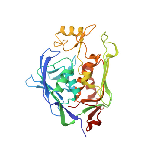 | |
UniProt | |||||
Entity Groups | |||||
| Sequence Clusters | 30% Identity50% Identity70% Identity90% Identity95% Identity100% Identity | ||||
| UniProt Group | P47205 | ||||
Sequence AnnotationsExpand | |||||
Reference Sequence | |||||
| Ligands 4 Unique | |||||
|---|---|---|---|---|---|
| ID | Chains | Name / Formula / InChI Key | 2D Diagram | 3D Interactions | |
| L63 Download:Ideal Coordinates CCD File | F [auth A], GA [auth D], O [auth B], Y [auth C] | N-[(2S,3R)-3-hydroxy-1-(hydroxyamino)-1-oxobutan-2-yl]biphenyl-4-carboxamide C17 H18 N2 O4 AXQUTBMFDJGETC-ABAIWWIYSA-N |  | ||
| GOL Download:Ideal Coordinates CCD File | AA [auth C] BA [auth C] CA [auth C] DA [auth C] G [auth A] | GLYCEROL C3 H8 O3 PEDCQBHIVMGVHV-UHFFFAOYSA-N |  | ||
| ZN Download:Ideal Coordinates CCD File | E [auth A], FA [auth D], N [auth B], X [auth C] | ZINC ION Zn PTFCDOFLOPIGGS-UHFFFAOYSA-N |  | ||
| ACT Download:Ideal Coordinates CCD File | EA [auth C] JA [auth D] K [auth A] L [auth A] M [auth B] | ACETATE ION C2 H3 O2 QTBSBXVTEAMEQO-UHFFFAOYSA-M |  | ||
| Length ( Å ) | Angle ( ˚ ) |
|---|---|
| a = 35.583 | α = 90 |
| b = 89.475 | β = 90.02 |
| c = 169.067 | γ = 90 |
| Software Name | Purpose |
|---|---|
| DENZO | data reduction |
| SCALEPACK | data scaling |
| BUSTER-TNT | refinement |
| PDB_EXTRACT | data extraction |
| MD2 | data collection |
| HKL-2000 | data reduction |
| PHASER | phasing |
| BUSTER | refinement |


