4FVV | pdb_00004fvv

Crystal structure of HCR/D-Sa-GBL1/C


Experimental Data Snapshot

  • Method: X-RAY DIFFRACTION
  • Resolution: 2.70 Å
  • R-Value Free: 
    0.263 (Depositor), 0.257 (DCC) 
  • R-Value Work: 
    0.206 (Depositor), 0.200 (DCC) 
  • R-Value Observed: 
    0.206 (Depositor) 

Starting Model: experimental
View more details

wwPDB Validation 3D Report Full Report

Validation slider image for 4FVV

Ligand Structure Quality Assessment 


This is version 1.3 of the entry. See complete history

Literature

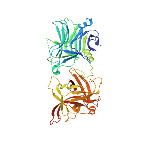
Botulinum neurotoxin serotype C associates with dual ganglioside receptors to facilitate cell entry.

Karalewitz, A.P.Fu, Z.Baldwin, M.R.Kim, J.J.Barbieri, J.T.

(2012) J Biological Chem 287: 40806-40816

  • DOI: https://doi.org/10.1074/jbc.M112.404244
  • Primary Citation Related Structures: 
    4FVV

  • PubMed Abstract: 

    How botulinum neurotoxin serotype C (BoNT/C) enters neurons is unclear. BoNT/C utilizes dual gangliosides as host cell receptors. BoNT/C accesses gangliosides on the plasma membrane. Plasma membrane accessibility of the dual ganglioside receptors suggests synaptic vesicle exocytosis may not be necessary to expose BoNT/C receptors. Botulinum neurotoxins (BoNTs) cleave SNARE proteins in motor neurons that inhibits synaptic vesicle (SV) exocytosis, resulting in flaccid paralysis. There are seven BoNT serotypes (A-G). In current models, BoNTs initially bind gangliosides on resting neurons and upon SV exocytosis associate with the luminal domains of SV-associated proteins as a second receptor. The entry of BoNT/C is less clear. Characterizing the heavy chain receptor binding domain (HCR), BoNT/C was shown to utilize gangliosides as dual host receptors. Crystallographic and biochemical studies showed that the two ganglioside binding sites, termed GBP2 and Sia-1, were independent and utilized unique mechanisms to bind complex gangliosides. The GBP2 binding site recognized gangliosides that contained a sia5 sialic acid, whereas the Sia-1 binding site recognized gangliosides that contained a sia7 sialic acid and sugars within the backbone of the ganglioside. Utilizing gangliosides that uniquely recognized the GBP2 and Sia-1 binding sites, HCR/C entry into Neuro-2A cells required both functional ganglioside binding sites. HCR/C entered cells differently than the HCR of tetanus toxin, which also utilizes dual gangliosides as host receptors. A point-mutated HCR/C that lacked GBP2 binding potential retained the ability to bind and enter Neuro-2A cells. This showed that ganglioside binding at the Sia-1 site was accessible on the plasma membrane, suggesting that SV exocytosis may not be required to expose BoNT/C receptors. These studies highlight the utility of BoNT HCRs as probes to study the role of gangliosides in neurotransmission.


  • Organizational Affiliation
    • Department of Microbiology and Molecular Genetics, Medical College of Wisconsin, Milwaukee, WI 53226, USA.

Macromolecule Content 

  • Total Structure Weight: 98.68 kDa 
  • Atom Count: 6,985 
  • Modeled Residue Count: 827 
  • Deposited Residue Count: 846 
  • Unique protein chains: 1

Macromolecules

Find similar proteins by:|  3D Structure
Entity ID: 1
MoleculeChains  Sequence LengthOrganismDetailsImage
Neurotoxin
A, B
423Clostridium botulinumMutation(s): 0 
UniProt
Find proteins for Q9LBR1 (Clostridium botulinum)
Explore Q9LBR1 
Go to UniProtKB:  Q9LBR1
Entity Groups
Sequence Clusters30% Identity50% Identity70% Identity90% Identity95% Identity100% Identity
UniProt GroupQ9LBR1
Sequence Annotations
Expand
Reference Sequence

Small Molecules

Ligands 3 Unique
IDChains Name / Formula / InChI Key2D Diagram3D Interactions
SIA

Query on SIA


Download:Ideal Coordinates CCD File
I [auth B]N-acetyl-alpha-neuraminic acid
C11 H19 N O9
SQVRNKJHWKZAKO-YRMXFSIDSA-N
SO4

Query on SO4


Download:Ideal Coordinates CCD File
C [auth A],
D [auth A],
E [auth A]
SULFATE ION
O4 S
QAOWNCQODCNURD-UHFFFAOYSA-L
GOL

Query on GOL


Download:Ideal Coordinates CCD File
F [auth A],
G [auth B],
H [auth B]
GLYCEROL
C3 H8 O3
PEDCQBHIVMGVHV-UHFFFAOYSA-N

Experimental Data & Validation

Experimental Data

  • Method: X-RAY DIFFRACTION
  • Resolution: 2.70 Å
  • R-Value Free:  0.263 (Depositor), 0.257 (DCC) 
  • R-Value Work:  0.206 (Depositor), 0.200 (DCC) 
  • R-Value Observed: 0.206 (Depositor) 
Space Group: I 2 2 2
Unit Cell:
Length ( Å )Angle ( ˚ )
a = 82.603α = 90
b = 154.63β = 90
c = 181.029γ = 90
Software Package:
Software NamePurpose
CrystalCleardata collection
PHASESphasing
CNSrefinement
HKL-2000data reduction
HKL-2000data scaling

Structure Validation

View Full Validation Report


Ligand Structure Quality Assessment 


Entry History 

Deposition Data

Revision History  (Full details and data files)

  • Version 1.0: 2012-10-24
    Type: Initial release
  • Version 1.1: 2012-12-12
    Changes: Database references
  • Version 1.2: 2020-07-29
    Type: Remediation
    Reason: Carbohydrate remediation
    Changes: Data collection, Derived calculations, Structure summary
  • Version 1.3: 2023-09-13
    Changes: Data collection, Database references, Refinement description, Structure summary