4FVK | pdb_00004fvk

Structural and functional characterization of neuraminidase-like molecule N10 derived from bat influenza A virus


Experimental Data Snapshot

  • Method: X-RAY DIFFRACTION
  • Resolution: 2.20 Å
  • R-Value Free: 
    0.246 (Depositor), 0.248 (DCC) 
  • R-Value Work: 
    0.207 (Depositor), 0.209 (DCC) 
  • R-Value Observed: 
    0.209 (Depositor) 

Starting Model: experimental
View more details

wwPDB Validation 3D Report Full Report

Validation slider image for 4FVK

Ligand Structure Quality Assessment 


This is version 2.2 of the entry. See complete history

Literature

Structural and functional characterization of neuraminidase-like molecule N10 derived from bat influenza A virus

Li, Q.Sun, X.M.Li, Z.X.Liu, Y.Vavricka, C.J.Qi, J.X.Gao, G.F.

(2012) Proc Natl Acad Sci U S A 109: 18897-18902

  • DOI: https://doi.org/10.1073/pnas.1211037109
  • Primary Citation Related Structures: 
    4FVK

  • PubMed Abstract: 

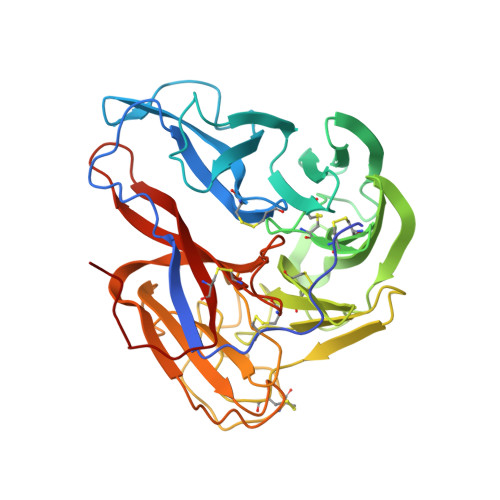
    The recent discovery of the unique genome of influenza virus H17N10 in bats raises considerable doubt about the origin and evolution of influenza A viruses. It also identifies a neuraminidase (NA)-like protein, N10, that is highly divergent from the nine other well-established serotypes of influenza A NA (N1-N9). The structural elucidation and functional characterization of influenza NAs have illustrated the complexity of NA structures, thus raising a key question as to whether N10 has a special structure and function. Here the crystal structure of N10, derived from influenza virus A/little yellow-shouldered bat/Guatemala/153/2009 (H17N10), was solved at a resolution of 2.20 Å. Overall, the structure of N10 was found to be similar to that of the other known influenza NA structures. In vitro enzymatic assays demonstrated that N10 lacks canonical NA activity. A detailed structural analysis revealed dramatic alterations of the conserved active site residues that are unfavorable for the binding and cleavage of terminally linked sialic acid receptors. Furthermore, an unusual 150-loop (residues 147-152) was observed to participate in the intermolecular polar interactions between adjacent N10 molecules of the N10 tetramer. Our study of influenza N10 provides insight into the structure and function of the sialidase superfamily and sheds light on the molecular mechanism of bat influenza virus infection.


  • Organizational Affiliation
    • School of Life Sciences, University of Science and Technology of China, Hefei 230027, China.

Macromolecule Content 

  • Total Structure Weight: 85.4 kDa 
  • Atom Count: 6,108 
  • Modeled Residue Count: 734 
  • Deposited Residue Count: 752 
  • Unique protein chains: 1

Macromolecules

Find similar proteins by:|  3D Structure
Entity ID: 1
MoleculeChains  Sequence LengthOrganismDetailsImage
Neuraminidase
A, B
376Influenza A virus (A/little yellow-shouldered bat/Guatemala/153/2009(H17N10))Mutation(s): 0 
Gene Names: NA
EC: 3.2.1.18
UniProt
Find proteins for H6QM75 (Influenza A virus (strain A/little yellow-shouldered bat/Guatemala/153/2009(H17N10)))
Explore H6QM75 
Go to UniProtKB:  H6QM75
Entity Groups
Sequence Clusters30% Identity50% Identity70% Identity90% Identity95% Identity100% Identity
UniProt GroupH6QM75
Glycosylation
Glycosylation Sites: 3
Sequence Annotations
Expand
Reference Sequence

Oligosaccharides

Help  
Entity ID: 2
MoleculeChains Length2D Diagram GlycosylationD Interactions
2-acetamido-2-deoxy-beta-D-glucopyranose-(1-4)-2-acetamido-2-deoxy-beta-D-glucopyranose
C
2N-Glycosylation
Glycosylation Resources
GlyTouCan: G42666HT
GlyCosmos: G42666HT
GlyGen: G42666HT

Small Molecules

Ligands 3 Unique
IDChains Name / Formula / InChI Key2D Diagram3D Interactions
NAG

Query on NAG


Download:Ideal Coordinates CCD File
H [auth A],
I [auth A],
N [auth B]
2-acetamido-2-deoxy-beta-D-glucopyranose
C8 H15 N O6
OVRNDRQMDRJTHS-FMDGEEDCSA-N
ZN

Query on ZN


Download:Ideal Coordinates CCD File
E [auth A]
F [auth A]
G [auth A]
K [auth B]
L [auth B]
E [auth A],
F [auth A],
G [auth A],
K [auth B],
L [auth B],
M [auth B]
ZINC ION
Zn
PTFCDOFLOPIGGS-UHFFFAOYSA-N
CA

Query on CA


Download:Ideal Coordinates CCD File
D [auth A],
J [auth B]
CALCIUM ION
Ca
BHPQYMZQTOCNFJ-UHFFFAOYSA-N

Experimental Data & Validation

Experimental Data

  • Method: X-RAY DIFFRACTION
  • Resolution: 2.20 Å
  • R-Value Free:  0.246 (Depositor), 0.248 (DCC) 
  • R-Value Work:  0.207 (Depositor), 0.209 (DCC) 
  • R-Value Observed: 0.209 (Depositor) 
Space Group: P 4 21 2
Unit Cell:
Length ( Å )Angle ( ˚ )
a = 122.496α = 90
b = 122.496β = 90
c = 110.504γ = 90
Software Package:
Software NamePurpose
Blu-Icedata collection
PHASERphasing
PHENIXrefinement
HKL-2000data reduction
HKL-2000data scaling

Structure Validation

View Full Validation Report


Ligand Structure Quality Assessment 


Entry History 

Deposition Data

Revision History  (Full details and data files)

  • Version 1.0: 2012-09-19
    Type: Initial release
  • Version 1.1: 2013-07-03
    Changes: Database references
  • Version 2.0: 2020-07-29
    Type: Remediation
    Reason: Carbohydrate remediation
    Changes: Atomic model, Data collection, Database references, Derived calculations, Structure summary
  • Version 2.1: 2023-11-08
    Changes: Data collection, Database references, Refinement description, Structure summary
  • Version 2.2: 2024-10-16
    Changes: Structure summary