4FU6 | pdb_00004fu6

Crystal structure of the PSIP1 PWWP domain


Experimental Data Snapshot

  • Method: X-RAY DIFFRACTION
  • Resolution: 2.10 Å
  • R-Value Free: 
    0.232 (Depositor), 0.242 (DCC) 
  • R-Value Work: 
    0.200 (Depositor), 0.210 (DCC) 
  • R-Value Observed: 
    0.201 (Depositor) 

Starting Model: experimental
View more details

wwPDB Validation 3D Report Full Report

Validation slider image for 4FU6

This is version 1.3 of the entry. See complete history

Literature

Structure and function of the nucleosome-binding PWWP domain.

Qin, S.Min, J.

(2014) Trends Biochem Sci 39: 536-547

  • DOI: https://doi.org/10.1016/j.tibs.2014.09.001
  • Primary Citation Related Structures: 
    4FU6

  • PubMed Abstract: 

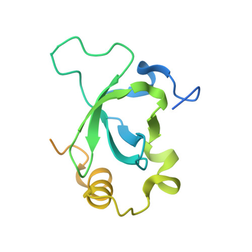
    PWWP domain-containing proteins are often involved in chromatin-associated biological processes, such as transcriptional regulation and DNA repair, and recent studies have shown that the PWWP domain specifies chromatin localization. Mutations in the PWWP domain, a 100-150 amino acid motif, have been linked to various human diseases, emphasizing its importance. Structural studies reveal that PWWP domains possess a conserved aromatic cage for histone methyl-lysine recognition, and synergistically bind both histone and DNA, which contributes to their nucleosome-binding ability and chromatin localization. Furthermore, the PWWP domain often cooperates with other histone and DNA 'reader' or 'modifier' domains to evoke crosstalk between various epigenetic marks. Here, we discuss these recent advances in understanding the structure and function of the PWWP domain.


  • Organizational Affiliation
    • Structural Genomics Consortium, University of Toronto, 101 College Street, Toronto, Ontario M5G 1L7, Canada.

Macromolecule Content 

  • Total Structure Weight: 17.9 kDa 
  • Atom Count: 808 
  • Modeled Residue Count: 95 
  • Deposited Residue Count: 153 
  • Unique protein chains: 1

Macromolecules

Find similar proteins by:|  3D Structure
Entity ID: 1
MoleculeChains  Sequence LengthOrganismDetailsImage
PC4 and SFRS1-interacting protein153Homo sapiensMutation(s): 0 
Gene Names: PSIP1DFS70LEDGFPSIP2
UniProt & NIH Common Fund Data Resources
Find proteins for O75475 (Homo sapiens)
Explore O75475 
Go to UniProtKB:  O75475
PHAROS:  O75475
GTEx:  ENSG00000164985 
Entity Groups
Sequence Clusters30% Identity50% Identity70% Identity90% Identity95% Identity100% Identity
UniProt GroupO75475
Sequence Annotations
Expand
Reference Sequence

Small Molecules

Ligands 3 Unique
IDChains Name / Formula / InChI Key2D Diagram3D Interactions
SO4

Query on SO4


Download:Ideal Coordinates CCD File
B [auth A],
C [auth A],
D [auth A]
SULFATE ION
O4 S
QAOWNCQODCNURD-UHFFFAOYSA-L
GOL

Query on GOL


Download:Ideal Coordinates CCD File
E [auth A]GLYCEROL
C3 H8 O3
PEDCQBHIVMGVHV-UHFFFAOYSA-N
UNX

Query on UNX


Download:Ideal Coordinates CCD File
F [auth A],
G [auth A],
H [auth A],
I [auth A],
J [auth A]
UNKNOWN ATOM OR ION
X

Experimental Data & Validation

Experimental Data

  • Method: X-RAY DIFFRACTION
  • Resolution: 2.10 Å
  • R-Value Free:  0.232 (Depositor), 0.242 (DCC) 
  • R-Value Work:  0.200 (Depositor), 0.210 (DCC) 
  • R-Value Observed: 0.201 (Depositor) 
Space Group: P 43 21 2
Unit Cell:
Length ( Å )Angle ( ˚ )
a = 45.96α = 90
b = 45.96β = 90
c = 156.24γ = 90
Software Package:
Software NamePurpose
DENZOdata reduction
SCALEPACKdata scaling
PHASERphasing
REFMACrefinement
PDB_EXTRACTdata extraction
HKL-2000data reduction
HKL-2000data scaling

Structure Validation

View Full Validation Report


Entry History 

Revision History  (Full details and data files)

  • Version 1.0: 2012-08-29
    Type: Initial release
  • Version 1.1: 2017-11-15
    Changes: Refinement description
  • Version 1.2: 2021-08-04
    Changes: Database references, Derived calculations
  • Version 1.3: 2023-09-13
    Changes: Data collection, Database references, Refinement description