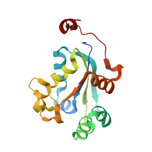
4FKX

Crystal structure of nucleoside diphosphate kinase B from Trypanosoma brucei bound to CDP


Experimental Data Snapshot

  • Method: X-RAY DIFFRACTION
  • Resolution: 1.70 Å
  • R-Value Free: 0.182 
  • R-Value Work: 0.152 
  • R-Value Observed: 0.154 

Starting Model: experimental
View more details

wwPDB Validation   3D Report Full Report


Ligand Structure Quality Assessment 


This is version 1.1 of the entry. See complete history


Literature

Crystal structure of nucleoside diphosphate kinase B from Trypanosoma brucei bound to CDP

Gardberg, A.S.Edwards, T.E.Seattle Structural Genomics Center for Infectious Disease (SSGCID)

To be published.

Macromolecules
Find similar proteins by:  (by identity cutoff)  |  3D Structure
Entity ID: 1
MoleculeChains Sequence LengthOrganismDetailsImage
Nucleoside diphosphate kinase
A, B, C
161Trypanosoma brucei brucei TREU927Mutation(s): 0 
Gene Names: Tb11.01.7800
EC: 2.7.4.6
UniProt
Find proteins for Q381H3 (Trypanosoma brucei brucei (strain 927/4 GUTat10.1))
Explore Q381H3 
Go to UniProtKB:  Q381H3
Entity Groups  
Sequence Clusters30% Identity50% Identity70% Identity90% Identity95% Identity100% Identity
UniProt GroupQ381H3
Sequence Annotations
Expand
  • Reference Sequence
Experimental Data & Validation

Experimental Data

Unit Cell:
Length ( Å )Angle ( ˚ )
a = 71.63α = 90
b = 121.37β = 90
c = 112.87γ = 90
Software Package:
Software NamePurpose
XSCALEdata scaling
PHASERphasing
REFMACrefinement
PDB_EXTRACTdata extraction
StructureStudiodata collection

Structure Validation

View Full Validation Report


Ligand Structure Quality Assessment 


Entry History 

Deposition Data

Revision History  (Full details and data files)

  • Version 1.0: 2012-06-27
    Type: Initial release
  • Version 1.1: 2023-09-13
    Changes: Data collection, Database references, Derived calculations, Refinement description