4FGC | pdb_00004fgc

Crystal Structure of Active Site Mutant C55A of Nitrile Reductase QueF, Bound to Substrate PreQ0


Experimental Data Snapshot

  • Method: X-RAY DIFFRACTION
  • Resolution: 2.50 Å
  • R-Value Free: 
    0.305 (Depositor), 0.298 (DCC) 
  • R-Value Work: 
    0.228 (Depositor), 0.233 (DCC) 
  • R-Value Observed: 
    0.232 (Depositor) 

Starting Model: experimental
View more details

wwPDB Validation 3D Report Full Report

Validation slider image for 4FGC

Ligand Structure Quality Assessment 


This is version 1.4 of the entry. See complete history

Literature

Structural basis of biological nitrile reduction.

Chikwana, V.M.Stec, B.Lee, B.W.de Crecy-Lagard, V.Iwata-Reuyl, D.Swairjo, M.A.

(2012) J Biological Chem 287: 30560-30570

  • DOI: https://doi.org/10.1074/jbc.M112.388538
  • Primary Citation Related Structures: 
    4F8B, 4FGC

  • PubMed Abstract: 

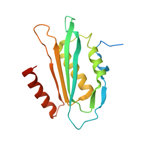
    The enzyme QueF catalyzes the reduction of the nitrile group of 7-cyano-7-deazaguanine (preQ(0)) to 7-aminomethyl-7-deazaguanine (preQ(1)), the only nitrile reduction reaction known in biology. We describe here two crystal structures of Bacillus subtilis QueF, one of the wild-type enzyme in complex with the substrate preQ(0), trapped as a covalent thioimide, a putative intermediate in the reaction, and the second of the C55A mutant in complex with the substrate preQ(0) bound noncovalently. The QueF enzyme forms an asymmetric tunnel-fold homodecamer of two head-to-head facing pentameric subunits, harboring 10 active sites at the intersubunit interfaces. In both structures, a preQ(0) molecule is bound at eight sites, and in the wild-type enzyme, it forms a thioimide covalent linkage to the catalytic residue Cys-55. Both structural and transient kinetic data show that preQ(0) binding, not thioimide formation, induces a large conformational change in and closure of the active site. Based on these data, we propose a mechanism for the activation of the Cys-55 nucleophile and subsequent hydride transfer.


  • Organizational Affiliation
    • Department of Chemistry, Portland State University, Portland, OR 97207, USA.

Macromolecule Content 

  • Total Structure Weight: 98.2 kDa 
  • Atom Count: 6,313 
  • Modeled Residue Count: 719 
  • Deposited Residue Count: 825 
  • Unique protein chains: 1

Macromolecules

Find similar proteins by:|  3D Structure
Entity ID: 1
MoleculeChains  Sequence LengthOrganismDetailsImage
NADPH-dependent 7-cyano-7-deazaguanine reductase
A, B, C, D, E
165Bacillus subtilis subsp. subtilis str. 168Mutation(s): 1 
Gene Names: BSU13750queFykvM
EC: 1.7.1.13
UniProt
Find proteins for O31678 (Bacillus subtilis (strain 168))
Explore O31678 
Go to UniProtKB:  O31678
Entity Groups
Sequence Clusters30% Identity50% Identity70% Identity90% Identity95% Identity100% Identity
UniProt GroupO31678
Sequence Annotations
Expand
Reference Sequence

Small Molecules

Ligands 3 Unique
IDChains Name / Formula / InChI Key2D Diagram3D Interactions
PE4

Query on PE4


Download:Ideal Coordinates CCD File
H [auth B]2-{2-[2-(2-{2-[2-(2-ETHOXY-ETHOXY)-ETHOXY]-ETHOXY}-ETHOXY)-ETHOXY]-ETHOXY}-ETHANOL
C16 H34 O8
PJWQOENWHPEPKI-UHFFFAOYSA-N
PQ0

Query on PQ0


Download:Ideal Coordinates CCD File
F [auth B],
J [auth C],
M [auth D],
P [auth E]
2-AMINO-4-OXO-4,7-DIHYDRO-3H-PYRROLO[2,3-D]PYRIMIDINE-5-CARBONITRILE
C7 H5 N5 O
FMKSMYDYKXQYRV-UHFFFAOYSA-N
CA

Query on CA


Download:Ideal Coordinates CCD File
G [auth B]
I [auth B]
K [auth C]
L [auth C]
N [auth D]
G [auth B],
I [auth B],
K [auth C],
L [auth C],
N [auth D],
O [auth D],
Q [auth E],
R [auth E]
CALCIUM ION
Ca
BHPQYMZQTOCNFJ-UHFFFAOYSA-N

Experimental Data & Validation

Experimental Data

  • Method: X-RAY DIFFRACTION
  • Resolution: 2.50 Å
  • R-Value Free:  0.305 (Depositor), 0.298 (DCC) 
  • R-Value Work:  0.228 (Depositor), 0.233 (DCC) 
  • R-Value Observed: 0.232 (Depositor) 
Space Group: P 32 2 1
Unit Cell:
Length ( Å )Angle ( ˚ )
a = 93.048α = 90
b = 93.048β = 90
c = 193.651γ = 120
Software Package:
Software NamePurpose
Blu-Icedata collection
REFMACrefinement
HKL-2000data reduction
HKL-2000data scaling
REFMACphasing

Structure Validation

View Full Validation Report


Ligand Structure Quality Assessment 


Entry History 

Deposition Data

Revision History  (Full details and data files)

  • Version 1.0: 2012-07-11
    Type: Initial release
  • Version 1.1: 2012-08-01
    Changes: Database references
  • Version 1.2: 2012-09-19
    Changes: Database references
  • Version 1.3: 2017-11-15
    Changes: Refinement description
  • Version 1.4: 2023-09-13
    Changes: Data collection, Database references, Derived calculations, Refinement description