4FCK | pdb_00004fck

Crystal Structure of the Co2+2-Human Arginase I-AGPA Complex


Experimental Data Snapshot

  • Method: X-RAY DIFFRACTION
  • Resolution: 1.90 Å
  • R-Value Free: 
    0.307 (Depositor), 0.305 (DCC) 
  • R-Value Work: 
    0.273 (Depositor), 0.273 (DCC) 
  • R-Value Observed: 
    0.273 (Depositor) 

Starting Model: experimental
View more details

wwPDB Validation 3D Report Full Report

Validation slider image for 4FCK

This is version 1.5 of the entry. See complete history

Literature

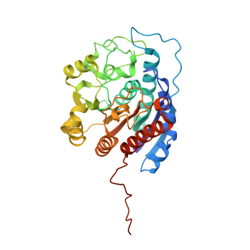
Binding of the unreactive substrate analog L-2-amino-3-guanidinopropionic acid (dinor-L-arginine) to human arginase I.

D'Antonio, E.L.Christianson, D.W.

(2012) Acta Crystallogr Sect F Struct Biol Cryst Commun 68: 889-893

  • DOI: https://doi.org/10.1107/S1744309112027820
  • Primary Citation Related Structures: 
    4FCI, 4FCK

  • PubMed Abstract: 

    Human arginase I (HAI) is a binuclear manganese metalloenzyme that catalyzes the hydrolysis of L-arginine to form L-ornithine and urea through a metal-activated hydroxide mechanism. Since HAI regulates L-Arg bioavailability for NO biosynthesis, it is a potential drug target for the treatment of cardiovascular diseases such as atherosclerosis. X-ray crystal structures are now reported of the complexes of Mn(2)(2+)-HAI and Co(2)(2+)-HAI with L-2-amino-3-guanidinopropionic acid (AGPA; also known as dinor-L-arginine), an amino acid bearing a guanidinium side chain two methylene groups shorter than that of L-arginine. Hydrogen bonds to the α-carboxylate and α-amino groups of AGPA dominate enzyme-inhibitor recognition; the guanidinium group does not interact directly with the metal ions.


  • Organizational Affiliation
    • Roy and Diana Vagelos Laboratories, Department of Chemistry, University of Pennsylvania, Philadelphia, PA 19104-6323, USA.

Macromolecule Content 

  • Total Structure Weight: 70.09 kDa 
  • Atom Count: 5,040 
  • Modeled Residue Count: 628 
  • Deposited Residue Count: 644 
  • Unique protein chains: 1

Macromolecules

Find similar proteins by:|  3D Structure
Entity ID: 1
MoleculeChains  Sequence LengthOrganismDetailsImage
Arginase-1
A, B
322Homo sapiensMutation(s): 0 
Gene Names: ARG1
EC: 3.5.3.1
UniProt & NIH Common Fund Data Resources
Find proteins for P05089 (Homo sapiens)
Explore P05089 
Go to UniProtKB:  P05089
PHAROS:  P05089
GTEx:  ENSG00000118520 
Entity Groups
Sequence Clusters30% Identity50% Identity70% Identity90% Identity95% Identity100% Identity
UniProt GroupP05089
Sequence Annotations
Expand
Reference Sequence

Experimental Data & Validation

Experimental Data

  • Method: X-RAY DIFFRACTION
  • Resolution: 1.90 Å
  • R-Value Free:  0.307 (Depositor), 0.305 (DCC) 
  • R-Value Work:  0.273 (Depositor), 0.273 (DCC) 
  • R-Value Observed: 0.273 (Depositor) 
Space Group: P 3
Unit Cell:
Length ( Å )Angle ( ˚ )
a = 90.451α = 90
b = 90.451β = 90
c = 69.362γ = 120
Software Package:
Software NamePurpose
CBASSdata collection
CNSrefinement
HKL-2000data reduction
HKL-2000data scaling
CNSphasing

Structure Validation

View Full Validation Report


Entry History 

Deposition Data

Revision History  (Full details and data files)

  • Version 1.0: 2012-06-20
    Type: Initial release
  • Version 1.1: 2012-08-15
    Changes: Database references
  • Version 1.2: 2013-01-23
    Changes: Database references
  • Version 1.3: 2018-12-05
    Changes: Data collection, Experimental preparation, Refinement description, Source and taxonomy
  • Version 1.4: 2019-11-20
    Changes: Derived calculations
  • Version 1.5: 2023-09-13
    Changes: Data collection, Database references, Derived calculations, Refinement description