4FC6 | pdb_00004fc6

Studies on DCR shed new light on peroxisomal beta-oxidation: Crystal structure of the ternary complex of pDCR


Experimental Data Snapshot

  • Method: X-RAY DIFFRACTION
  • Resolution: 2.10 Å
  • R-Value Free: 
    0.215 (Depositor), 0.214 (DCC) 
  • R-Value Work: 
    0.156 (Depositor), 0.156 (DCC) 
  • R-Value Observed: 
    0.159 (Depositor) 

wwPDB Validation 3D Report Full Report

Validation slider image for 4FC6

Ligand Structure Quality Assessment 


This is version 1.3 of the entry. See complete history

Literature

Studies of human 2,4-dienoyl CoA reductase shed new light on peroxisomal beta-oxidation of unsaturated fatty acids

Hua, T.Wu, D.Ding, W.Wang, J.Shaw, N.Liu, Z.J.

(2012) J Biological Chem 287: 28956-28965

  • DOI: https://doi.org/10.1074/jbc.M112.385351
  • Primary Citation Related Structures: 
    4FC6, 4FC7

  • PubMed Abstract: 

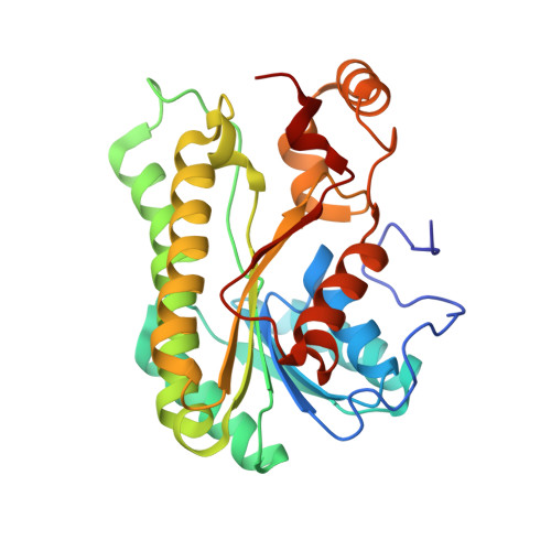
    Peroxisomes play an essential role in maintaining fatty acid homeostasis. Although mitochondria are also known to participate in the catabolism of fatty acids via β-oxidation, differences exist between the peroxisomal and mitochondrial β-oxidation. Only peroxisomes, but not mitochondrion, can shorten very long chain fatty acids. Here, we describe the crystal structure of a ternary complex of peroxisomal 2,4-dienoyl CoA reductases (pDCR) with hexadienoyl CoA and NADP, as a prototype for comparison with the mitochondrial 2,4-dienoyl CoA reductase (mDCR) to shed light on the differences between the enzymes from the two organelles at the molecular level. Unexpectedly, the structure of pDCR refined to 1.84 Å resolution reveals the absence of the tyrosine-serine pair seen in the active site of mDCR, which together with a lysine and an asparagine have been deemed a hallmark of the SDR family of enzymes. Instead, aspartate hydrogen-bonded to the Cα hydroxyl via a water molecule seems to perturb the water molecule for protonation of the substrate. Our studies provide the first structural evidence for participation of water in the DCR-catalyzed reactions. Biochemical studies and structural analysis suggest that pDCRs can catalyze the shortening of six-carbon-long substrates in vitro. However, the K(m) values of pDCR for short chain acyl CoAs are at least 6-fold higher than those for substrates with 10 or more aliphatic carbons. Unlike mDCR, hinge movements permit pDCR to process very long chain polyunsaturated fatty acids.


  • Organizational Affiliation
    • National Laboratory of Biomacromolecules, Institute of Biophysics, Chinese Academy of Sciences, Beijing 100101, China.

Macromolecule Content 

  • Total Structure Weight: 123.31 kDa 
  • Atom Count: 9,358 
  • Modeled Residue Count: 1,098 
  • Deposited Residue Count: 1,108 
  • Unique protein chains: 1

Macromolecules

Find similar proteins by:|  3D Structure
Entity ID: 1
MoleculeChains  Sequence LengthOrganismDetailsImage
Peroxisomal 2,4-dienoyl-CoA reductase
A, B, C, D
277Homo sapiensMutation(s): 0 
Gene Names: DECR2PDCR
EC: 1.3.1.34 (PDB Primary Data), 1.3.1.124 (UniProt)
UniProt & NIH Common Fund Data Resources
Find proteins for Q9NUI1 (Homo sapiens)
Explore Q9NUI1 
Go to UniProtKB:  Q9NUI1
PHAROS:  Q9NUI1
GTEx:  ENSG00000242612 
Entity Groups
Sequence Clusters30% Identity50% Identity70% Identity90% Identity95% Identity100% Identity
UniProt GroupQ9NUI1
Sequence Annotations
Expand
Reference Sequence

Small Molecules

Ligands 2 Unique
IDChains Name / Formula / InChI Key2D Diagram3D Interactions
HXC

Query on HXC


Download:Ideal Coordinates CCD File
F [auth A],
H [auth B],
I [auth C],
L [auth D]
HEXANOYL-COENZYME A
C27 H46 N7 O17 P3 S
OEXFMSFODMQEPE-HDRQGHTBSA-N
NAP

Query on NAP


Download:Ideal Coordinates CCD File
E [auth A],
G [auth B],
J [auth C],
K [auth D]
NADP NICOTINAMIDE-ADENINE-DINUCLEOTIDE PHOSPHATE
C21 H28 N7 O17 P3
XJLXINKUBYWONI-NNYOXOHSSA-N

Experimental Data & Validation

Experimental Data

  • Method: X-RAY DIFFRACTION
  • Resolution: 2.10 Å
  • R-Value Free:  0.215 (Depositor), 0.214 (DCC) 
  • R-Value Work:  0.156 (Depositor), 0.156 (DCC) 
  • R-Value Observed: 0.159 (Depositor) 
Space Group: P 21 21 21
Unit Cell:
Length ( Å )Angle ( ˚ )
a = 88.038α = 90
b = 95.114β = 90
c = 133.409γ = 90
Software Package:
Software NamePurpose
HKL-2000data collection
PHASESphasing
REFMACrefinement
HKL-2000data reduction
HKL-2000data scaling

Structure Validation

View Full Validation Report


Ligand Structure Quality Assessment 


Entry History 

Deposition Data

Revision History  (Full details and data files)

  • Version 1.0: 2012-07-04
    Type: Initial release
  • Version 1.1: 2012-07-11
    Changes: Database references
  • Version 1.2: 2013-07-24
    Changes: Database references
  • Version 1.3: 2024-02-28
    Changes: Data collection, Database references, Derived calculations