4FB8 | pdb_00004fb8

Crystal Structure of apo Acyl-CoA Carboxylase


Experimental Data Snapshot

  • Method: X-RAY DIFFRACTION
  • Resolution: 3.00 Å
  • R-Value Free: 
    0.304 (Depositor), 0.293 (DCC) 
  • R-Value Work: 
    0.233 (Depositor), 0.233 (DCC) 
  • R-Value Observed: 
    0.236 (Depositor) 

Starting Model: experimental
View more details

wwPDB Validation 3D Report Full Report

Validation slider image for 4FB8

This is version 1.4 of the entry. See complete history

Literature

Structure, Activity, and Inhibition of the Carboxyltransferase beta-Subunit of Acetyl Coenzyme A Carboxylase (AccD6) from Mycobacterium tuberculosis.

Reddy, M.C.Breda, A.Bruning, J.B.Sherekar, M.Valluru, S.Thurman, C.Ehrenfeld, H.Sacchettini, J.C.

(2014) Antimicrob Agents Chemother 58: 6122-6132

  • DOI: https://doi.org/10.1128/AAC.02574-13
  • Primary Citation Related Structures: 
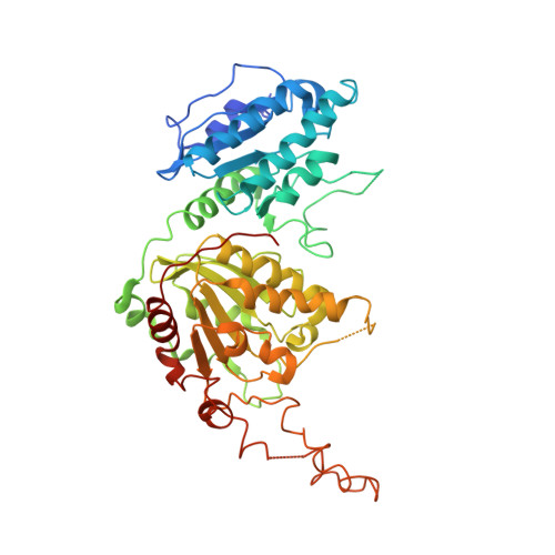
    4FB8, 4G2R

  • PubMed Abstract: 

    In Mycobacterium tuberculosis, the carboxylation of acetyl coenzyme A (acetyl-CoA) to produce malonyl-CoA, a building block in long-chain fatty acid biosynthesis, is catalyzed by two enzymes working sequentially: a biotin carboxylase (AccA) and a carboxyltransferase (AccD). While the exact roles of the three different biotin carboxylases (AccA1 to -3) and the six carboxyltransferases (AccD1 to -6) in M. tuberculosis are still not clear, AccD6 in complex with AccA3 can synthesize malonyl-CoA from acetyl-CoA. A series of 10 herbicides that target plant acetyl-CoA carboxylases (ACC) were tested for inhibition of AccD6 and for whole-cell activity against M. tuberculosis. From the tested herbicides, haloxyfop, an arylophenoxypropionate, showed in vitro inhibition of M. tuberculosis AccD6, with a 50% inhibitory concentration (IC50) of 21.4 ± 1 μM. Here, we report the crystal structures of M. tuberculosis AccD6 in the apo form (3.0 Å) and in complex with haloxyfop-R (2.3 Å). The structure of M. tuberculosis AccD6 in complex with haloxyfop-R shows two molecules of the inhibitor bound on each AccD6 subunit. These results indicate the potential for developing novel therapeutics for tuberculosis based on herbicides with low human toxicity.


  • Organizational Affiliation
    • Department of Biochemistry and Biophysics, Texas A&M University, College Station, Texas, USA.

Macromolecule Content 

  • Total Structure Weight: 100.4 kDa 
  • Atom Count: 6,220 
  • Modeled Residue Count: 862 
  • Deposited Residue Count: 946 
  • Unique protein chains: 1

Macromolecules

Find similar proteins by:|  3D Structure
Entity ID: 1
MoleculeChains  Sequence LengthOrganismDetailsImage
Probable propionyl-CoA carboxylase beta chain 6
A, B
473Mycobacterium tuberculosisMutation(s): 0 
Gene Names: accD6MT2307MTCY427.28Rv2247
EC: 6.4.1.3 (PDB Primary Data), 2.1.3.15 (UniProt), 2.1.3 (UniProt)
UniProt
Find proteins for P9WQH5 (Mycobacterium tuberculosis (strain ATCC 25618 / H37Rv))
Explore P9WQH5 
Go to UniProtKB:  P9WQH5
Entity Groups
Sequence Clusters30% Identity50% Identity70% Identity90% Identity95% Identity100% Identity
UniProt GroupP9WQH5
Sequence Annotations
Expand
Reference Sequence

Experimental Data & Validation

Experimental Data

  • Method: X-RAY DIFFRACTION
  • Resolution: 3.00 Å
  • R-Value Free:  0.304 (Depositor), 0.293 (DCC) 
  • R-Value Work:  0.233 (Depositor), 0.233 (DCC) 
  • R-Value Observed: 0.236 (Depositor) 
Space Group: P 21 21 21
Unit Cell:
Length ( Å )Angle ( ˚ )
a = 82.284α = 90
b = 82.361β = 90
c = 157.87γ = 90
Software Package:
Software NamePurpose
DENZOdata reduction
SCALEPACKdata scaling
PHASERphasing
PHENIXrefinement
PDB_EXTRACTdata extraction

Structure Validation

View Full Validation Report


Entry History 

Revision History  (Full details and data files)

  • Version 1.0: 2014-02-19
    Type: Initial release
  • Version 1.1: 2014-08-13
    Changes: Database references
  • Version 1.2: 2014-10-01
    Changes: Database references
  • Version 1.3: 2017-11-15
    Changes: Refinement description
  • Version 1.4: 2023-09-13
    Changes: Data collection, Database references, Refinement description