4F8V

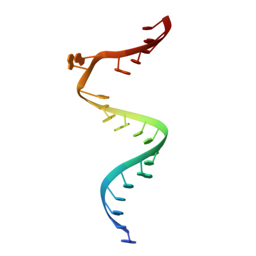
Crystal structure of the bacterial ribosomal decoding site in complex with sisomicin (P21212 form)


Experimental Data Snapshot

  • Method: X-RAY DIFFRACTION
  • Resolution: 2.80 Å
  • R-Value Free: 0.265 
  • R-Value Work: 0.226 

wwPDB Validation   3D Report Full Report


Ligand Structure Quality Assessment 


This is version 1.3 of the entry. See complete history


Literature

Crystal structure and specific binding mode of sisomicin to the bacterial ribosomal decoding site.

Kondo, J.Koganei, M.Kasahara, T.

(2012) ACS Med Chem Lett 3: 741-744

  • DOI: https://doi.org/10.1021/ml300145y
  • Primary Citation of Related Structures:  
    4F8U, 4F8V

  • PubMed Abstract: 

    Sisomicin with an unsaturated sugar ring I displays better antibacterial activity than other structurally related aminoglycosides, such as gentamicin, tobramycin, and amikacin. In the present study, we have confirmed by X-ray analyses that the binding mode of sisomicin is basically similar but not identical to that of the related compounds having saturated ring I. A remarkable difference is found in the stacking interaction between ring I and G1491. While the typical saturated ring I with a chair conformation stacks on G1491 through CH/π interactions, the unsaturated ring I of sisomicin with a partially planar conformation can share its π-electron density with G1491 and fits well within the A-site helix.


  • Organizational Affiliation

    Department of Materials and Life Sciences, Faculty of Science and Technology, Sophia University , 7-1 Kioi-cho, Chiyoda-ku, 102-8554 Tokyo, Japan.


Macromolecules

Find similar nucleic acids by:  Sequence   |   3D Structure  

Entity ID: 1
MoleculeChains LengthOrganismImage
RNA (5'-R(P*GP*CP*GP*UP*CP*AP*CP*AP*CP*CP*GP*GP*UP*GP*AP*AP*GP*UP*CP*GP*C)-3')
A, B
21N/A
Sequence Annotations
Expand
  • Reference Sequence
Small Molecules
Ligands 1 Unique
IDChains Name / Formula / InChI Key2D Diagram3D Interactions
SIS
Query on SIS

Download Ideal Coordinates CCD File 
C [auth A],
D [auth B]
(1S,2S,3R,4S,6R)-4,6-diamino-3-{[(2S,3R)-3-amino-6-(aminomethyl)-3,4-dihydro-2H-pyran-2-yl]oxy}-2-hydroxycyclohexyl 3-deoxy-4-C-methyl-3-(methylamino)-beta-L-arabinopyranoside
C19 H37 N5 O7
URWAJWIAIPFPJE-YFMIWBNJSA-N
Experimental Data & Validation

Experimental Data

  • Method: X-RAY DIFFRACTION
  • Resolution: 2.80 Å
  • R-Value Free: 0.265 
  • R-Value Work: 0.226 
  • Space Group: P 21 21 2
Unit Cell:
Length ( Å )Angle ( ˚ )
a = 31.65α = 90
b = 107.65β = 90
c = 48.86γ = 90
Software Package:
Software NamePurpose
ADSCdata collection
PHENIXmodel building
CNSrefinement
CrystalCleardata reduction
CrystalCleardata scaling
PHENIXphasing

Structure Validation

View Full Validation Report


Ligand Structure Quality Assessment 


Entry History 

Deposition Data

Revision History  (Full details and data files)

  • Version 1.0: 2012-08-15
    Type: Initial release
  • Version 1.1: 2012-09-05
    Changes: Structure summary
  • Version 1.2: 2019-12-25
    Changes: Database references
  • Version 1.3: 2024-03-20
    Changes: Data collection, Database references, Derived calculations