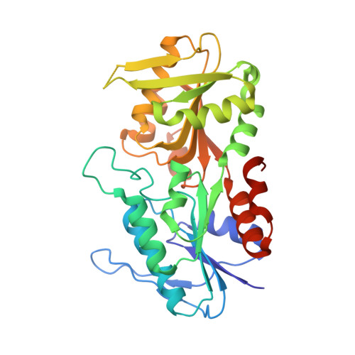
4F8E

Crystal structure of human PRS1 D52H mutant


Experimental Data Snapshot

  • Method: X-RAY DIFFRACTION
  • Resolution: 2.27 Å
  • R-Value Free: 0.233 
  • R-Value Work: 0.178 

wwPDB Validation   3D Report Full Report


This is version 1.2 of the entry. See complete history


Literature

A small disturbance, but a serious disease: the possible mechanism of D52H-mutant of human PRS1 that causes gout

Chen, P.Li, J.Ma, J.Teng, M.Li, X.

(2013) IUBMB Life 65: 518-525

  • DOI: https://doi.org/10.1002/iub.1154
  • Primary Citation of Related Structures:  
    4F8E

  • PubMed Abstract: 

    Phosphoribosyl pyrophosphate synthetase isoform 1 (PRS1) has an essential role in the de novo and salvage synthesis of human purine and pyrimidine nucleotides. The dysfunction of PRS1 will dramatically influence nucleotides' concentration in patient's body and lead to different kinds of disorders (such as hyperuricemia, gout and deafness). The D52H missense mutation of PRS1 will lead to a conspicuous phosphoribosyl pyrophosphate content elevation in the erythrocyte of patients and finally induce hyperuricemia and serious gout. In this study, the enzyme activity analysis indicated that D52H-mutant possessed similar catalytic activity to the wild-type PRS1, and the 2.27 Å resolution D52H-mutant crystal structure revealed that the stable interaction network surrounding the 52 position of PRS1 would be completely destroyed by the substitution of histidine. These interaction variations would further influence the conformation of ADP-binding pocket of D52H-mutant and reduced the inhibitor sensitivity of PRS1 in patient's body.


  • Organizational Affiliation

    University of Science and Technology of China, School of Life Sciences, Hefei, Anhui, People's Republic of China.


Macromolecules
Find similar proteins by:  (by identity cutoff)  |  3D Structure
Entity ID: 1
MoleculeChains Sequence LengthOrganismDetailsImage
Ribose-phosphate pyrophosphokinase 1
A, B
326Homo sapiensMutation(s): 1 
Gene Names: PRPS1
EC: 2.7.6.1
UniProt & NIH Common Fund Data Resources
Find proteins for P60891 (Homo sapiens)
Explore P60891 
Go to UniProtKB:  P60891
PHAROS:  P60891
GTEx:  ENSG00000147224 
Entity Groups  
Sequence Clusters30% Identity50% Identity70% Identity90% Identity95% Identity100% Identity
UniProt GroupP60891
Sequence Annotations
Expand
  • Reference Sequence
Small Molecules
Ligands 2 Unique
IDChains Name / Formula / InChI Key2D Diagram3D Interactions
SO4
Query on SO4

Download Ideal Coordinates CCD File 
C [auth A]
D [auth A]
E [auth A]
G [auth B]
H [auth B]
C [auth A],
D [auth A],
E [auth A],
G [auth B],
H [auth B],
I [auth B]
SULFATE ION
O4 S
QAOWNCQODCNURD-UHFFFAOYSA-L
MG
Query on MG

Download Ideal Coordinates CCD File 
F [auth A],
J [auth B]
MAGNESIUM ION
Mg
JLVVSXFLKOJNIY-UHFFFAOYSA-N
Experimental Data & Validation

Experimental Data

  • Method: X-RAY DIFFRACTION
  • Resolution: 2.27 Å
  • R-Value Free: 0.233 
  • R-Value Work: 0.178 
  • Space Group: H 3
Unit Cell:
Length ( Å )Angle ( ˚ )
a = 170.31α = 90
b = 170.31β = 90
c = 61.75γ = 120
Software Package:
Software NamePurpose
HKL-2000data collection
MOLREPphasing
REFMACrefinement
HKL-2000data reduction
HKL-2000data scaling

Structure Validation

View Full Validation Report


Entry History 

Deposition Data

Revision History  (Full details and data files)

  • Version 1.0: 2013-05-22
    Type: Initial release
  • Version 1.1: 2014-03-12
    Changes: Database references
  • Version 1.2: 2024-03-20
    Changes: Data collection, Database references, Derived calculations