4F7R | pdb_00004f7r

Crystal structure of 14-3-3 protein from Giardia intestinalis


Experimental Data Snapshot

  • Method: X-RAY DIFFRACTION
  • Resolution: 3.20 Å
  • R-Value Free: 
    0.232 (Depositor), 0.232 (DCC) 
  • R-Value Work: 
    0.185 (Depositor), 0.192 (DCC) 
  • R-Value Observed: 
    0.187 (Depositor) 

wwPDB Validation 3D Report Full Report

Validation slider image for 4F7R

This is version 1.3 of the entry. See complete history

Literature

The Crystal Structure of Giardia duodenalis 14-3-3 in the Apo Form: When Protein Post-Translational Modifications Make the Difference.

Fiorillo, A.di Marino, D.Bertuccini, L.Via, A.Pozio, E.Camerini, S.Ilari, A.Lalle, M.

(2014) PLoS One 9: e92902-e92902

  • DOI: https://doi.org/10.1371/journal.pone.0092902
  • Primary Citation Related Structures: 
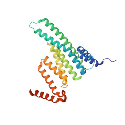
    4F7R

  • PubMed Abstract: 

    The 14-3-3s are a family of dimeric evolutionary conserved pSer/pThr binding proteins that play a key role in multiple biological processes by interacting with a plethora of client proteins. Giardia duodenalis is a flagellated protozoan that affects millions of people worldwide causing an acute and chronic diarrheal disease. The single giardial 14-3-3 isoform (g14-3-3), unique in the 14-3-3 family, needs the constitutive phosphorylation of Thr214 and the polyglycylation of its C-terminus to be fully functional in vivo. Alteration of the phosphorylation and polyglycylation status affects the parasite differentiation into the cyst stage. To further investigate the role of these post-translational modifications, the crystal structure of the g14-3-3 was solved in the unmodified apo form. Oligomers of g14-3-3 were observed due to domain swapping events at the protein C-terminus. The formation of filaments was supported by TEM. Mutational analysis, in combination with native PAGE and chemical cross-linking, proved that polyglycylation prevents oligomerization. In silico phosphorylation and molecular dynamics simulations supported a structural role for the phosphorylation of Thr214 in promoting target binding. Our findings highlight unique structural features of g14-3-3 opening novel perspectives on the evolutionary history of this protein family and envisaging the possibility to develop anti-giardial drugs targeting g14-3-3.


  • Organizational Affiliation
    • Department of Biochemical Sciences "A. Rossi-Fanelli", University of Rome "Sapienza", Rome, Italy; Institute of Molecular Biology and Pathology, CNR, Rome, Italy and Institute Pasteur Cenci-Bolognetti Foundation at Department of Biochemical Sciences "A. Rossi-Fanelli", University of Rome "Sapienza", Rome, Italy.

Macromolecule Content 

  • Total Structure Weight: 117.63 kDa 
  • Atom Count: 7,801 
  • Modeled Residue Count: 948 
  • Deposited Residue Count: 1,012 
  • Unique protein chains: 1

Macromolecules

Find similar proteins by:|  3D Structure
Entity ID: 1
MoleculeChains  Sequence LengthOrganismDetailsImage
14-3-3 protein
A, B, C, D
253Giardia duodenalisMutation(s): 0 
UniProt
Find proteins for E2RU97 (Giardia intestinalis (strain ATCC 50803 / WB clone C6))
Explore E2RU97 
Go to UniProtKB:  E2RU97
Entity Groups
Sequence Clusters30% Identity50% Identity70% Identity90% Identity95% Identity100% Identity
UniProt GroupE2RU97
Sequence Annotations
Expand
Reference Sequence

Small Molecules

Ligands 1 Unique
IDChains Name / Formula / InChI Key2D Diagram3D Interactions
SO4

Query on SO4


Download:Ideal Coordinates CCD File
E [auth A]
F [auth A]
G [auth A]
H [auth A]
I [auth A]
E [auth A],
F [auth A],
G [auth A],
H [auth A],
I [auth A],
J [auth B],
K [auth B],
L [auth B],
M [auth B],
N [auth C],
O [auth C],
P [auth C],
Q [auth C],
R [auth C],
S [auth D],
T [auth D]
SULFATE ION
O4 S
QAOWNCQODCNURD-UHFFFAOYSA-L

Experimental Data & Validation

Experimental Data

  • Method: X-RAY DIFFRACTION
  • Resolution: 3.20 Å
  • R-Value Free:  0.232 (Depositor), 0.232 (DCC) 
  • R-Value Work:  0.185 (Depositor), 0.192 (DCC) 
  • R-Value Observed: 0.187 (Depositor) 
Space Group: P 32
Unit Cell:
Length ( Å )Angle ( ˚ )
a = 100.879α = 90
b = 100.879β = 90
c = 140.559γ = 120
Software Package:
Software NamePurpose
ADSCdata collection
PHASERphasing
REFMACrefinement
DENZOdata reduction
SCALEPACKdata scaling

Structure Validation

View Full Validation Report


Entry History 

Deposition Data

Revision History  (Full details and data files)

  • Version 1.0: 2013-05-22
    Type: Initial release
  • Version 1.1: 2014-04-16
    Changes: Database references
  • Version 1.2: 2015-04-01
    Changes: Structure summary
  • Version 1.3: 2024-02-28
    Changes: Data collection, Database references, Derived calculations, Refinement description