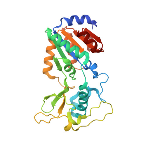
4F56

The bicyclic intermediate structure provides insights into the desuccinylation mechanism of SIRT5


Experimental Data Snapshot

  • Method: X-RAY DIFFRACTION
  • Resolution: 1.70 Å
  • R-Value Free: 0.233 
  • R-Value Work: 0.169 
  • R-Value Observed: 0.172 

Starting Model: experimental
View more details

wwPDB Validation   3D Report Full Report


Ligand Structure Quality Assessment 


This is version 1.4 of the entry. See complete history


Literature

The Bicyclic Intermediate Structure Provides Insights into the Desuccinylation Mechanism of Human Sirtuin 5 (SIRT5)

Zhou, Y.Zhang, H.He, B.Du, J.Lin, H.Cerione, R.A.Hao, Q.

(2012) J Biol Chem 287: 28307-28314

  • DOI: https://doi.org/10.1074/jbc.M112.384511
  • Primary Citation of Related Structures:  
    4F4U, 4F56

  • PubMed Abstract: 

    Sirtuins are pivotal regulators in various cellular processes, including transcription, DNA repair, genome stability, and energy metabolism. Their functions have been generally attributed to NAD-dependent deacetylase activity. However, human SIRT5 (sirtuin 5), which has been reported to exhibit little deacetylase activity, was recently identified as an NAD-dependent demalonylase and desuccinylase. Biochemical studies suggested that the mechanism of SIRT5-catalyzed demalonylation and desuccinylation is similar to that of deacetylation catalyzed by other sirtuins. Previously, we solved the crystal structure of a SIRT5-succinyl-lysine peptide-NAD complex. Here, we present two more structures: a binary complex of SIRT5 with an H3K9 succinyl peptide and a binary complex of SIRT5 with a bicyclic intermediate obtained by incubating SIRT5-H3K9 thiosuccinyl peptide co-crystals with NAD. To our knowledge, this represents the first bicyclic intermediate for a sirtuin-catalyzed deacylation reaction that has been captured in a crystal structure, thus providing unique insights into the reaction mechanism. The structural information should benefit the design of specific inhibitors for SIRT5 and help in exploring the therapeutic potential of targeting sirtuins for treating human diseases.


  • Organizational Affiliation

    MacCHESS, Cornell High Energy Synchrotron Source, Cornell University, Ithaca, New York 14853, USA.


Macromolecules
Find similar proteins by:  (by identity cutoff)  |  3D Structure
Entity ID: 1
MoleculeChains Sequence LengthOrganismDetailsImage
NAD-dependent lysine demalonylase and desuccinylase sirtuin-5, mitochondrial
A, B
273Homo sapiensMutation(s): 0 
Gene Names: SIR2L5SIRT5
EC: 3.5.1 (PDB Primary Data), 2.3.1 (UniProt)
UniProt & NIH Common Fund Data Resources
Find proteins for Q9NXA8 (Homo sapiens)
Explore Q9NXA8 
Go to UniProtKB:  Q9NXA8
PHAROS:  Q9NXA8
GTEx:  ENSG00000124523 
Entity Groups  
Sequence Clusters30% Identity50% Identity70% Identity90% Identity95% Identity100% Identity
UniProt GroupQ9NXA8
Sequence Annotations
Expand
  • Reference Sequence

Find similar proteins by:  Sequence   |   3D Structure  

Entity ID: 2
MoleculeChains Sequence LengthOrganismDetailsImage
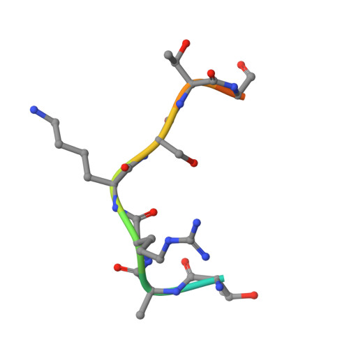
peptide from Histone H3.1
C, D
12Homo sapiensMutation(s): 0 
UniProt & NIH Common Fund Data Resources
Find proteins for P68431 (Homo sapiens)
Explore P68431 
Go to UniProtKB:  P68431
PHAROS:  P68431
Entity Groups  
Sequence Clusters30% Identity50% Identity70% Identity90% Identity95% Identity100% Identity
UniProt GroupP68431
Sequence Annotations
Expand
  • Reference Sequence
Small Molecules
Ligands 2 Unique
IDChains Name / Formula / InChI Key2D Diagram3D Interactions
CGK
Query on CGK

Download Ideal Coordinates CCD File 
G [auth C],
H [auth D]
3-[(2R,3aR,5R,6R,6aR)-5-({[(S)-{[(S)-{[(2R,3S,4R,5R)-5-(6-amino-9H-purin-9-yl)-3,4-dihydroxytetrahydrofuran-2-yl]methoxy}(hydroxy)phosphoryl]oxy}(hydroxy)phosphoryl]oxy}methyl)-2,6-dihydroxytetrahydrofuro[2,3-d][1,3]oxathiol-2-yl]propanoic acid
C19 H27 N5 O16 P2 S
DLPJYEOHCQEPAR-IOFPOCHNSA-N
ZN
Query on ZN

Download Ideal Coordinates CCD File 
E [auth A],
F [auth B]
ZINC ION
Zn
PTFCDOFLOPIGGS-UHFFFAOYSA-N
Experimental Data & Validation

Experimental Data

  • Method: X-RAY DIFFRACTION
  • Resolution: 1.70 Å
  • R-Value Free: 0.233 
  • R-Value Work: 0.169 
  • R-Value Observed: 0.172 
  • Space Group: P 21 21 21
Unit Cell:
Length ( Å )Angle ( ˚ )
a = 52.403α = 90
b = 66.755β = 90
c = 156.861γ = 90
Software Package:
Software NamePurpose
DENZOdata reduction
SCALEPACKdata scaling
REFMACrefinement
PDB_EXTRACTdata extraction
CrystalCleardata collection
HKL-2000data reduction
MOLREPphasing

Structure Validation

View Full Validation Report


Ligand Structure Quality Assessment 


Entry History 

Deposition Data

Revision History  (Full details and data files)

  • Version 1.0: 2012-06-20
    Type: Initial release
  • Version 1.1: 2012-09-19
    Changes: Database references
  • Version 1.2: 2017-11-15
    Changes: Refinement description
  • Version 1.3: 2023-11-08
    Changes: Data collection, Database references, Derived calculations, Refinement description
  • Version 1.4: 2024-10-16
    Changes: Structure summary