4F4Q | pdb_00004f4q

Crystal structure of M. smegmatis DprE1 in complex with FAD and covalently bound BTZ043


Experimental Data Snapshot

  • Method: X-RAY DIFFRACTION
  • Resolution: 2.62 Å
  • R-Value Free: 
    0.240 (Depositor), 0.240 (DCC) 
  • R-Value Work: 
    0.199 (Depositor), 0.197 (DCC) 
  • R-Value Observed: 
    0.201 (Depositor) 

Starting Model: experimental
View more details

wwPDB Validation 3D Report Full Report

Validation slider image for 4F4Q

Ligand Structure Quality Assessment 


This is version 1.5 of the entry. See complete history

Literature

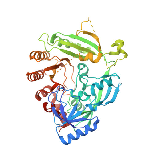
Structural Basis for Benzothiazinone-Mediated Killing of Mycobacterium tuberculosis.

Neres, J.Pojer, F.Molteni, E.Chiarelli, L.R.Dhar, N.Boy-Rottger, S.Buroni, S.Fullam, E.Degiacomi, G.Lucarelli, A.P.Read, R.J.Zanoni, G.Edmondson, D.E.De Rossi, E.Pasca, M.R.McKinney, J.D.Dyson, P.J.Riccardi, G.Mattevi, A.Cole, S.T.Binda, C.

(2012) Sci Transl Med 4: 150ra121-150ra121

  • DOI: https://doi.org/10.1126/scitranslmed.3004395
  • Primary Citation Related Structures: 
    4AUT, 4F4Q

  • PubMed Abstract: 

    The benzothiazinone BTZ043 is a tuberculosis drug candidate with nanomolar whole-cell activity. BTZ043 targets the DprE1 catalytic component of the essential enzyme decaprenylphosphoryl-β-D-ribofuranose-2'-epimerase, thus blocking biosynthesis of arabinans, vital components of mycobacterial cell walls. Crystal structures of DprE1, in its native form and in a complex with BTZ043, reveal formation of a semimercaptal adduct between the drug and an active-site cysteine, as well as contacts to a neighboring catalytic lysine residue. Kinetic studies confirm that BTZ043 is a mechanism-based, covalent inhibitor. This explains the exquisite potency of BTZ043, which, when fluorescently labeled, localizes DprE1 at the poles of growing bacteria. Menaquinone can reoxidize the flavin adenine dinucleotide cofactor in DprE1 and may be the natural electron acceptor for this reaction in the mycobacterium. Our structural and kinetic analysis provides both insight into a critical epimerization reaction and a platform for structure-based design of improved inhibitors.


  • Organizational Affiliation
    • Global Health Institute, École Polytechnique Fédérale de Lausanne, CH-1015 Lausanne, Switzerland.

Macromolecule Content 

  • Total Structure Weight: 52.49 kDa 
  • Atom Count: 3,358 
  • Modeled Residue Count: 412 
  • Deposited Residue Count: 468 
  • Unique protein chains: 1

Macromolecules

Find similar proteins by:|  3D Structure
Entity ID: 1
MoleculeChains  Sequence LengthOrganismDetailsImage
DprE1468Mycolicibacterium smegmatis MC2 155Mutation(s): 0 
Gene Names: MSMEG_6382
EC: 1.1.98.3
UniProt
Find proteins for A0R607 (Mycolicibacterium smegmatis (strain ATCC 700084 / mc(2)155))
Explore A0R607 
Go to UniProtKB:  A0R607
Entity Groups
Sequence Clusters30% Identity50% Identity70% Identity90% Identity95% Identity100% Identity
UniProt GroupA0R607
Sequence Annotations
Expand
Reference Sequence

Small Molecules

Ligands 3 Unique
IDChains Name / Formula / InChI Key2D Diagram3D Interactions
FAD

Query on FAD


Download:Ideal Coordinates CCD File
C [auth A]FLAVIN-ADENINE DINUCLEOTIDE
C27 H33 N9 O15 P2
VWWQXMAJTJZDQX-UYBVJOGSSA-N
0SK

Query on 0SK


Download:Ideal Coordinates CCD File
B [auth A]8-(hydroxyamino)-2-[(2S)-2-methyl-1,4-dioxa-8-azaspiro[4.5]dec-8-yl]-6-(trifluoromethyl)-4H-1,3-benzothiazin-4-one
C17 H18 F3 N3 O4 S
WTTODOLGMIMGNK-VIFPVBQESA-N
GOL

Query on GOL


Download:Ideal Coordinates CCD File
D [auth A]GLYCEROL
C3 H8 O3
PEDCQBHIVMGVHV-UHFFFAOYSA-N

Experimental Data & Validation

Experimental Data

  • Method: X-RAY DIFFRACTION
  • Resolution: 2.62 Å
  • R-Value Free:  0.240 (Depositor), 0.240 (DCC) 
  • R-Value Work:  0.199 (Depositor), 0.197 (DCC) 
  • R-Value Observed: 0.201 (Depositor) 
Space Group: P 21 21 21
Unit Cell:
Length ( Å )Angle ( ˚ )
a = 39.924α = 90
b = 64.309β = 90
c = 202.841γ = 90
Software Package:
Software NamePurpose
PHASERphasing
REFMACrefinement
XDSdata reduction
XDSdata scaling

Structure Validation

View Full Validation Report


Ligand Structure Quality Assessment 


Entry History 

Deposition Data

Revision History  (Full details and data files)

  • Version 1.0: 2012-09-05
    Type: Initial release
  • Version 1.1: 2012-09-12
    Changes: Database references
  • Version 1.2: 2012-09-19
    Changes: Database references
  • Version 1.3: 2017-11-15
    Changes: Refinement description
  • Version 1.4: 2023-09-13
    Changes: Data collection, Database references, Derived calculations, Refinement description
  • Version 1.5: 2024-11-27
    Changes: Structure summary