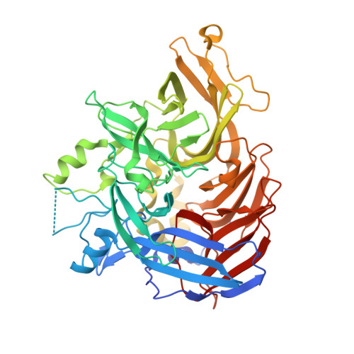
4F3A

Structure of RPE65: P6522 crystal form, iridium derivative


Experimental Data Snapshot

  • Method: X-RAY DIFFRACTION
  • Resolution: 2.60 Å
  • R-Value Free: 0.229 
  • R-Value Work: 0.185 
  • R-Value Observed: 0.187 

wwPDB Validation   3D Report Full Report


This is version 1.3 of the entry. See complete history


Literature

Structure of RPE65 isomerase in a lipidic matrix reveals roles for phospholipids and iron in catalysis.

Kiser, P.D.Farquhar, E.R.Shi, W.Sui, X.Chance, M.R.Palczewski, K.

(2012) Proc Natl Acad Sci U S A 109: E2747-E2756

  • DOI: https://doi.org/10.1073/pnas.1212025109
  • Primary Citation of Related Structures:  
    4F2Z, 4F30, 4F3A, 4F3D

  • PubMed Abstract: 

    RPE65 is a key metalloenzyme responsible for maintaining visual function in vertebrates. Despite extensive research on this membrane-bound retinoid isomerase, fundamental questions regarding its enzymology remain unanswered. Here, we report the crystal structure of RPE65 in a membrane-like environment. These crystals, obtained from enzymatically active, nondelipidated protein, displayed an unusual packing arrangement wherein RPE65 is embedded in a lipid-detergent sheet. Structural differences between delipidated and nondelipidated RPE65 uncovered key residues involved in substrate uptake and processing. Complementary iron K-edge X-ray absorption spectroscopy data established that RPE65 as isolated contained a divalent iron center and demonstrated the presence of a tightly bound ligand consistent with a coordinated carboxylate group. These results support the hypothesis that the Lewis acidity of iron could be used to promote ester dissociation and generation of a carbocation intermediate required for retinoid isomerization.


  • Organizational Affiliation

    Department of Pharmacology, School of Medicine, Case Western Reserve University, Cleveland, OH 44106, USA. pdk7@case.edu


Macromolecules
Find similar proteins by:  (by identity cutoff)  |  3D Structure
Entity ID: 1
MoleculeChains Sequence LengthOrganismDetailsImage
Retinoid isomerohydrolase533Bos taurusMutation(s): 0 
EC: 3.1.1.64 (PDB Primary Data), 5.3.3.22 (UniProt)
Membrane Entity: Yes 
UniProt
Find proteins for Q28175 (Bos taurus)
Explore Q28175 
Go to UniProtKB:  Q28175
Entity Groups  
Sequence Clusters30% Identity50% Identity70% Identity90% Identity95% Identity100% Identity
UniProt GroupQ28175
Sequence Annotations
Expand
  • Reference Sequence
Small Molecules
Ligands 2 Unique
IDChains Name / Formula / InChI Key2D Diagram3D Interactions
IR3
Query on IR3

Download Ideal Coordinates CCD File 
B [auth A]IRIDIUM (III) ION
Ir
MILUBEOXRNEUHS-UHFFFAOYSA-N
FE2
Query on FE2

Download Ideal Coordinates CCD File 
C [auth A]FE (II) ION
Fe
CWYNVVGOOAEACU-UHFFFAOYSA-N
Experimental Data & Validation

Experimental Data

  • Method: X-RAY DIFFRACTION
  • Resolution: 2.60 Å
  • R-Value Free: 0.229 
  • R-Value Work: 0.185 
  • R-Value Observed: 0.187 
  • Space Group: P 65 2 2
Unit Cell:
Length ( Å )Angle ( ˚ )
a = 178.639α = 90
b = 178.639β = 90
c = 86.469γ = 120
Software Package:
Software NamePurpose
XDSdata scaling
SHELXCDphasing
SHELXEmodel building
PHASERphasing
ARP/wARPmodel building
REFMACrefinement
XDSdata reduction

Structure Validation

View Full Validation Report


Entry History 

Deposition Data

Revision History  (Full details and data files)

  • Version 1.0: 2012-10-03
    Type: Initial release
  • Version 1.1: 2014-07-02
    Changes: Database references
  • Version 1.2: 2017-11-15
    Changes: Refinement description
  • Version 1.3: 2024-02-28
    Changes: Data collection, Database references, Derived calculations