4F2O | pdb_00004f2o

Quisqualate bound to the D655A mutant of the ligand binding domain of GluA3


Experimental Data Snapshot

  • Method: X-RAY DIFFRACTION
  • Resolution: 1.91 Å
  • R-Value Free: 
    0.228 (Depositor), 0.222 (DCC) 
  • R-Value Work: 
    0.188 (Depositor), 0.186 (DCC) 
  • R-Value Observed: 
    0.192 (Depositor) 

Starting Model: experimental
View more details

wwPDB Validation 3D Report Full Report

Validation slider image for 4F2O

Ligand Structure Quality Assessment 


This is version 1.3 of the entry. See complete history

Literature

The loss of an electrostatic contact unique to AMPA receptor ligand binding domain 2 slows channel activation.

Holley, S.M.Ahmed, A.H.Srinivasan, J.Murthy, S.E.Weiland, G.A.Oswald, R.E.Nowak, L.M.

(2012) Biochemistry 51: 4015-4027

  • DOI: https://doi.org/10.1021/bi3001837
  • Primary Citation Related Structures: 
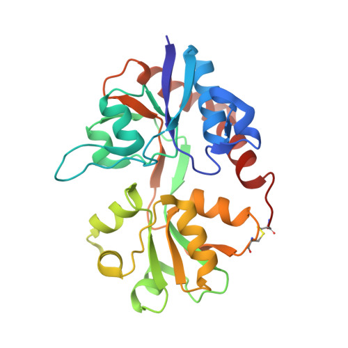
    4F1Y, 4F22, 4F29, 4F2O, 4F2Q, 4F31, 4F39, 4F3B, 4F3G

  • PubMed Abstract: 

    Ligand-gated ion channels undergo conformational changes that transfer the energy of agonist binding to channel opening. Within ionotropic glutamate receptor (iGluR) subunits, this process is initiated in their bilobate ligand binding domain (LBD) where agonist binding to lobe 1 favors closure of lobe 2 around the agonist and allows formation of interlobe hydrogen bonds. AMPA receptors (GluAs) differ from other iGluRs because glutamate binding causes an aspartate-serine peptide bond in a flexible part of lobe 2 to rotate 180° (flipped conformation), allowing these residues to form cross-cleft H-bonds with tyrosine and glycine in lobe 1. This aspartate also contacts the side chain of a lysine residue in the hydrophobic core of lobe 2 by a salt bridge. We investigated how the peptide flip and electrostatic contact (D655-K660) in GluA3 contribute to receptor function by examining pharmacological and structural properties with an antagonist (CNQX), a partial agonist (kainate), and two full agonists (glutamate and quisqualate) in the wildtype and two mutant receptors. Alanine substitution decreased the agonist potency of GluA3(i)-D655A and GluA3(i)-K660A receptor channels expressed in HEK293 cells and differentially affected agonist binding affinity for isolated LBDs without changing CNQX affinity. Correlations observed in the crystal structures of the mutant LBDs included the loss of the D655-K660 electrostatic contact, agonist-dependent differences in lobe 1 and lobe 2 closure, and unflipped D(A)655-S656 bonds. Glutamate-stimulated activation was slower for both mutants, suggesting that efficient energy transfer of agonist binding within the LBD of AMPA receptors requires an intact tether between the flexible peptide flip domain and the rigid hydrophobic core of lobe 2.


  • Organizational Affiliation
    • Department of Molecular Medicine, Cornell University, Ithaca, NY 14853, USA.

Macromolecule Content 

  • Total Structure Weight: 29.18 kDa 
  • Atom Count: 2,390 
  • Modeled Residue Count: 258 
  • Deposited Residue Count: 258 
  • Unique protein chains: 1

Macromolecules

Find similar proteins by:|  3D Structure
Entity ID: 1
MoleculeChains  Sequence LengthOrganismDetailsImage
Glutamate receptor 3258Rattus norvegicusMutation(s): 1 
Gene Names: Glur3Gria3
UniProt
Find proteins for P19492 (Rattus norvegicus)
Explore P19492 
Go to UniProtKB:  P19492
Entity Groups
Sequence Clusters30% Identity50% Identity70% Identity90% Identity95% Identity100% Identity
UniProt GroupP19492
Sequence Annotations
Expand
Reference Sequence

Small Molecules

Ligands 2 Unique
IDChains Name / Formula / InChI Key2D Diagram3D Interactions
QUS

Query on QUS


Download:Ideal Coordinates CCD File
B [auth A](S)-2-AMINO-3-(3,5-DIOXO-[1,2,4]OXADIAZOLIDIN-2-YL)-PROPIONIC ACID
C5 H7 N3 O5
ASNFTDCKZKHJSW-REOHCLBHSA-N
ZN

Query on ZN


Download:Ideal Coordinates CCD File
C [auth A]ZINC ION
Zn
PTFCDOFLOPIGGS-UHFFFAOYSA-N

Experimental Data & Validation

Experimental Data

  • Method: X-RAY DIFFRACTION
  • Resolution: 1.91 Å
  • R-Value Free:  0.228 (Depositor), 0.222 (DCC) 
  • R-Value Work:  0.188 (Depositor), 0.186 (DCC) 
  • R-Value Observed: 0.192 (Depositor) 
Space Group: P 2 2 21
Unit Cell:
Length ( Å )Angle ( ˚ )
a = 47.594α = 90
b = 47.825β = 90
c = 136.555γ = 90
Software Package:
Software NamePurpose
HKL-3000data collection
PHENIXmodel building
PHENIXrefinement
HKL-3000data reduction
HKL-3000data scaling
PHENIXphasing

Structure Validation

View Full Validation Report


Ligand Structure Quality Assessment 


Entry History 

Deposition Data

Revision History  (Full details and data files)

  • Version 1.0: 2012-05-23
    Type: Initial release
  • Version 1.1: 2017-08-16
    Changes: Data collection, Source and taxonomy
  • Version 1.2: 2023-09-13
    Changes: Data collection, Database references, Derived calculations, Refinement description
  • Version 1.3: 2024-11-06
    Changes: Structure summary