


Crystal Structure of Escherichia coli L-arabinose Isomerase (ECAI) complexed with Ribitol
Manjasetty, B.A., Burley, S.K., Almo, S.C., Chance, M.R.To be published.
Experimental Data Snapshot
Starting Model: experimental
View more details
Entity ID: 1 | |||||
|---|---|---|---|---|---|
| Molecule | Chains | Sequence Length | Organism | Details | Image |
| L-arabinose isomerase | 500 | Escherichia coli K-12 | Mutation(s): 0 Gene Names: araA, b0062, JW0061, UTI89_C0067 EC: 5.3.1.4 | 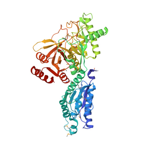 | |
UniProt | |||||
Entity Groups | |||||
| Sequence Clusters | 30% Identity50% Identity70% Identity90% Identity95% Identity100% Identity | ||||
| UniProt Group | P08202 | ||||
Sequence AnnotationsExpand | |||||
Reference Sequence | |||||
| Ligands 3 Unique | |||||
|---|---|---|---|---|---|
| ID | Chains | Name / Formula / InChI Key | 2D Diagram | 3D Interactions | |
| RB0 Download:Ideal Coordinates CCD File | E [auth A], G [auth B], J [auth C] | D-ribitol C5 H12 O5 HEBKCHPVOIAQTA-ZXFHETKHSA-N |  | ||
| ACY Download:Ideal Coordinates CCD File | H [auth B] | ACETIC ACID C2 H4 O2 QTBSBXVTEAMEQO-UHFFFAOYSA-N |  | ||
| MN Download:Ideal Coordinates CCD File | D [auth A], F [auth B], I [auth C] | MANGANESE (II) ION Mn WAEMQWOKJMHJLA-UHFFFAOYSA-N |  | ||
| Modified Residues 1 Unique | |||||
|---|---|---|---|---|---|
| ID | Chains | Type | Formula | 2D Diagram | Parent |
| MSE Query on MSE | A, B, C | L-PEPTIDE LINKING | C5 H11 N O2 Se |  | MET |
| Length ( Å ) | Angle ( ˚ ) |
|---|---|
| a = 116.472 | α = 90 |
| b = 116.472 | β = 90 |
| c = 214.81 | γ = 120 |
| Software Name | Purpose |
|---|---|
| CBASS | data collection |
| MOLREP | phasing |
| REFMAC | refinement |
| HKL-2000 | data reduction |
| SCALEPACK | data scaling |


