4EY5 | pdb_00004ey5

Crystal Structure of Recombinant Human Acetylcholinesterase in Complex with (-)-huperzine A


Experimental Data Snapshot

  • Method: X-RAY DIFFRACTION
  • Resolution: 2.30 Å
  • R-Value Free: 
    0.212 (Depositor), 0.213 (DCC) 
  • R-Value Work: 
    0.176 (Depositor), 0.180 (DCC) 
  • R-Value Observed: 
    0.177 (Depositor) 

Starting Model: experimental
View more details

wwPDB Validation 3D Report Full Report

Validation slider image for 4EY5

Ligand Structure Quality Assessment 


This is version 2.2 of the entry. See complete history

Literature

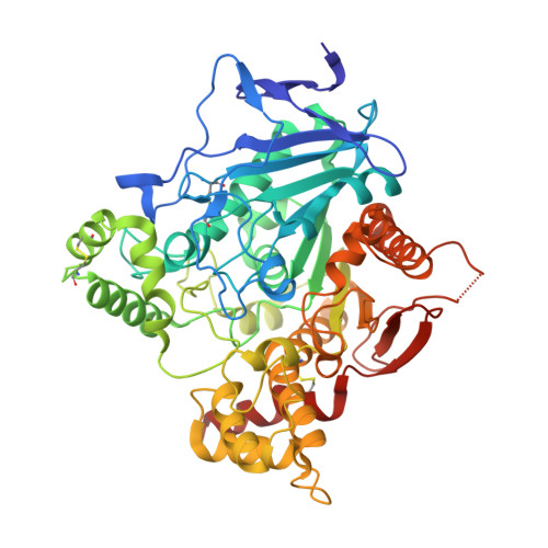
Structures of human acetylcholinesterase in complex with pharmacologically important ligands.

Cheung, J.Rudolph, M.J.Burshteyn, F.Cassidy, M.S.Gary, E.N.Love, J.Franklin, M.C.Height, J.J.

(2012) J Med Chem 55: 10282-10286

  • DOI: https://doi.org/10.1021/jm300871x
  • Primary Citation Related Structures: 
    4EY4, 4EY5, 4EY6, 4EY7, 4EY8

  • PubMed Abstract: 

    Human acetylcholinesterase (AChE) is a significant target for therapeutic drugs. Here we present high resolution crystal structures of human AChE, alone and in complexes with drug ligands; donepezil, an Alzheimer's disease drug, binds differently to human AChE than it does to Torpedo AChE. These crystals of human AChE provide a more accurate platform for further drug development than previously available.


  • Organizational Affiliation
    • New York Structural Biology Center, New York, New York 10027, USA.

Macromolecule Content 

  • Total Structure Weight: 121.21 kDa 
  • Atom Count: 8,878 
  • Modeled Residue Count: 1,065 
  • Deposited Residue Count: 1,084 
  • Unique protein chains: 1

Macromolecules

Find similar proteins by:|  3D Structure
Entity ID: 1
MoleculeChains  Sequence LengthOrganismDetailsImage
Acetylcholinesterase
A, B
542Homo sapiensMutation(s): 0 
Gene Names: ACHE
EC: 3.1.1.7
UniProt & NIH Common Fund Data Resources
Find proteins for P22303 (Homo sapiens)
Explore P22303 
Go to UniProtKB:  P22303
PHAROS:  P22303
GTEx:  ENSG00000087085 
Entity Groups
Sequence Clusters30% Identity50% Identity70% Identity90% Identity95% Identity100% Identity
UniProt GroupP22303
Glycosylation
Glycosylation Sites: 2Go to GlyGen: P22303-1
Sequence Annotations
Expand
Reference Sequence

Oligosaccharides

Help  
Entity ID: 2
MoleculeChains Length2D Diagram GlycosylationD Interactions
2-acetamido-2-deoxy-beta-D-glucopyranose-(1-4)-[alpha-L-fucopyranose-(1-6)]2-acetamido-2-deoxy-beta-D-glucopyranose
C, D
3N-Glycosylation
Glycosylation Resources
GlyTouCan: G21290RB
GlyCosmos: G21290RB
GlyGen: G21290RB

Small Molecules

Ligands 5 Unique
IDChains Name / Formula / InChI Key2D Diagram3D Interactions
HUP

Query on HUP


Download:Ideal Coordinates CCD File
E [auth A],
J [auth B]
Huperzine A
C15 H18 N2 O
ZRJBHWIHUMBLCN-YQEJDHNASA-N
NAG

Query on NAG


Download:Ideal Coordinates CCD File
I [auth B]2-acetamido-2-deoxy-beta-D-glucopyranose
C8 H15 N O6
OVRNDRQMDRJTHS-FMDGEEDCSA-N
DMS

Query on DMS


Download:Ideal Coordinates CCD File
F [auth A],
K [auth B]
DIMETHYL SULFOXIDE
C2 H6 O S
IAZDPXIOMUYVGZ-UHFFFAOYSA-N
EDO

Query on EDO


Download:Ideal Coordinates CCD File
G [auth A],
H [auth A],
L [auth B]
1,2-ETHANEDIOL
C2 H6 O2
LYCAIKOWRPUZTN-UHFFFAOYSA-N
NO3

Query on NO3


Download:Ideal Coordinates CCD File
M [auth B],
N [auth B]
NITRATE ION
N O3
NHNBFGGVMKEFGY-UHFFFAOYSA-N
Binding Affinity Annotations 
IDSourceBinding Affinity
HUP BindingDB:  4EY5 Kd: 17 (nM) from 1 assay(s)
IC50: min: 21, max: 5280 (nM) from 19 assay(s)

Experimental Data & Validation

Experimental Data

  • Method: X-RAY DIFFRACTION
  • Resolution: 2.30 Å
  • R-Value Free:  0.212 (Depositor), 0.213 (DCC) 
  • R-Value Work:  0.176 (Depositor), 0.180 (DCC) 
  • R-Value Observed: 0.177 (Depositor) 
Space Group: P 31 2 1
Unit Cell:
Length ( Å )Angle ( ˚ )
a = 105.222α = 90
b = 105.222β = 90
c = 323.289γ = 120
Software Package:
Software NamePurpose
DENZOdata reduction
SCALEPACKdata scaling
PHENIXrefinement
PDB_EXTRACTdata extraction
HKL-2000data collection
HKL-2000data reduction
HKL-2000data scaling

Structure Validation

View Full Validation Report


Ligand Structure Quality Assessment 


Entry History 

Deposition Data

Revision History  (Full details and data files)

  • Version 1.0: 2012-10-17
    Type: Initial release
  • Version 1.1: 2012-12-12
    Changes: Database references
  • Version 1.2: 2013-10-23
    Changes: Non-polymer description
  • Version 1.3: 2017-11-15
    Changes: Refinement description
  • Version 2.0: 2020-07-29
    Type: Remediation
    Reason: Carbohydrate remediation
    Changes: Atomic model, Data collection, Derived calculations, Structure summary
  • Version 2.1: 2023-09-13
    Changes: Data collection, Database references, Refinement description, Structure summary
  • Version 2.2: 2024-11-06
    Changes: Structure summary