4ESQ | pdb_00004esq

Crystal structure of the extracellular domain of PknH from Mycobacterium tuberculosis


Experimental Data Snapshot

  • Method: X-RAY DIFFRACTION
  • Resolution: 1.70 Å
  • R-Value Free: 
    0.197 (Depositor), 0.197 (DCC) 
  • R-Value Work: 
    0.163 (Depositor), 0.161 (DCC) 
  • R-Value Observed: 
    0.165 (Depositor) 

wwPDB Validation 3D Report Full Report

Validation slider image for 4ESQ

This is version 1.3 of the entry. See complete history

Literature

Structure of the sensor domain of Mycobacterium tuberculosis PknH receptor kinase reveals a conserved binding cleft.

Cavazos, A.Prigozhin, D.M.Alber, T.

(2012) J Mol Biology 422: 488-494

  • DOI: https://doi.org/10.1016/j.jmb.2012.06.011
  • Primary Citation Related Structures: 
    4ESQ

  • PubMed Abstract: 

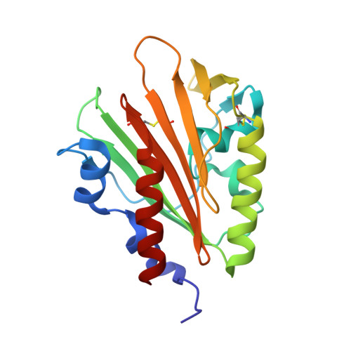
    Since their discovery over 20 years ago, eukaryotic-like transmembrane receptor Ser/Thr protein kinases (STPKs) have been shown to play critical roles in the virulence, growth, persistence, and reactivation of many bacteria. Information regarding the signals transmitted by these proteins, however, remains scarce. To enhance understanding of the basis for STPK receptor signaling, we determined the 1.7-Å-resolution crystal structure of the extracellular sensor domain of the Mycobacterium tuberculosis receptor STPK, PknH (Rv1266c). The PknH sensor domain adopts an unanticipated fold containing two intramolecular disulfide bonds and a large hydrophobic and polar cleft. The residues lining the cleft and those surrounding the disulfide bonds are conserved. These results suggest that PknH binds a small-molecule ligand that signals by changing the location or quaternary structure of the kinase domain.


  • Organizational Affiliation
    • Department of Molecular and Cell Biology, University of California, Berkeley, CA 94720, USA.

Macromolecule Content 

  • Total Structure Weight: 21.19 kDa 
  • Atom Count: 1,704 
  • Modeled Residue Count: 194 
  • Deposited Residue Count: 194 
  • Unique protein chains: 1

Macromolecules

Find similar proteins by:|  3D Structure
Entity ID: 1
MoleculeChains  Sequence LengthOrganismDetailsImage
Serine/threonine protein kinase194Mycobacterium tuberculosis H37RvMutation(s): 0 
Gene Names: pknHRv1266c
EC: 2.7.11.1
UniProt
Find proteins for P9WI71 (Mycobacterium tuberculosis (strain ATCC 25618 / H37Rv))
Explore P9WI71 
Go to UniProtKB:  P9WI71
Entity Groups
Sequence Clusters30% Identity50% Identity70% Identity90% Identity95% Identity100% Identity
UniProt GroupP9WI71
Sequence Annotations
Expand
Reference Sequence

Experimental Data & Validation

Experimental Data

  • Method: X-RAY DIFFRACTION
  • Resolution: 1.70 Å
  • R-Value Free:  0.197 (Depositor), 0.197 (DCC) 
  • R-Value Work:  0.163 (Depositor), 0.161 (DCC) 
  • R-Value Observed: 0.165 (Depositor) 
Space Group: P 1 21 1
Unit Cell:
Length ( Å )Angle ( ˚ )
a = 47.455α = 90
b = 35.923β = 98.36
c = 49.308γ = 90
Software Package:
Software NamePurpose
DENZOdata reduction
SCALEPACKdata scaling
SOLVEphasing
RESOLVEphasing
PHENIXrefinement
PDB_EXTRACTdata extraction

Structure Validation

View Full Validation Report


Entry History 

Deposition Data

Revision History  (Full details and data files)

  • Version 1.0: 2012-07-18
    Type: Initial release
  • Version 1.1: 2013-01-02
    Changes: Database references
  • Version 1.2: 2017-11-15
    Changes: Refinement description
  • Version 1.3: 2024-11-20
    Changes: Data collection, Database references, Derived calculations, Structure summary