4EOH

Crystal Structure of Human PL Kinase with bound Theophylline


Experimental Data Snapshot

  • Method: X-RAY DIFFRACTION
  • Resolution: 2.10 Å
  • R-Value Free: 0.254 
  • R-Value Work: 0.205 

wwPDB Validation   3D Report Full Report


Ligand Structure Quality Assessment 


This is version 1.2 of the entry. See complete history


Literature

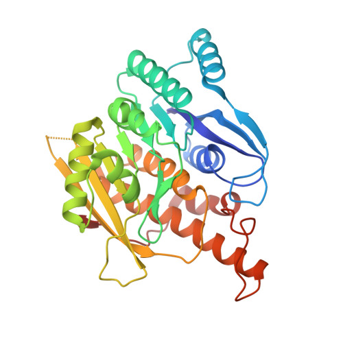
Crystal structures of human pyridoxal kinase in complex with the neurotoxins, ginkgotoxin and theophylline: insights into pyridoxal kinase inhibition.

Gandhi, A.K.Desai, J.V.Ghatge, M.S.di Salvo, M.L.Di Biase, S.Danso-Danquah, R.Musayev, F.N.Contestabile, R.Schirch, V.Safo, M.K.

(2012) PLoS One 7: e40954-e40954

  • DOI: https://doi.org/10.1371/journal.pone.0040954
  • Primary Citation of Related Structures:  
    4EN4, 4EOH

  • PubMed Abstract: 

    Several drugs and natural compounds are known to be highly neurotoxic, triggering epileptic convulsions or seizures, and causing headaches, agitations, as well as other neuronal symptoms. The neurotoxic effects of some of these compounds, including theophylline and ginkgotoxin, have been traced to their inhibitory activity against human pyridoxal kinase (hPL kinase), resulting in deficiency of the active cofactor form of vitamin B₆, pyridoxal 5'-phosphate (PLP). Pyridoxal (PL), an inactive form of vitamin B₆ is converted to PLP by PL kinase. PLP is the B₆ vitamer required as a cofactor for over 160 enzymatic activities essential in primary and secondary metabolism. We have performed structural and kinetic studies on hPL kinase with several potential inhibitors, including ginkgotoxin and theophylline. The structural studies show ginkgotoxin and theophylline bound at the substrate site, and are involved in similar protein interactions as the natural substrate, PL. Interestingly, the phosphorylated product of ginkgotoxin is also observed bound at the active site. This work provides insights into the molecular basis of hPL kinase inhibition and may provide a working hypothesis to quickly screen or identify neurotoxic drugs as potential hPL kinase inhibitors. Such adverse effects may be prevented by administration of an appropriate form of vitamin B₆, or provide clues of how to modify these drugs to help reduce their hPL kinase inhibitory effects.


  • Organizational Affiliation

    Department of Medicinal Chemistry, Institute for Structural Biology and Drug Discovery, Virginia Commonwealth University, Richmond, Virginia, United States of America.


Macromolecules
Find similar proteins by:  (by identity cutoff)  |  3D Structure
Entity ID: 1
MoleculeChains Sequence LengthOrganismDetailsImage
Pyridoxal Kinase
A, B
312Homo sapiensMutation(s): 0 
Gene Names: PDXKC21orf124C21orf97PKHPNKPRED79
EC: 2.7.1.35
UniProt & NIH Common Fund Data Resources
Find proteins for O00764 (Homo sapiens)
Explore O00764 
Go to UniProtKB:  O00764
PHAROS:  O00764
GTEx:  ENSG00000160209 
Entity Groups  
Sequence Clusters30% Identity50% Identity70% Identity90% Identity95% Identity100% Identity
UniProt GroupO00764
Sequence Annotations
Expand
  • Reference Sequence
Small Molecules
Ligands 4 Unique
IDChains Name / Formula / InChI Key2D Diagram3D Interactions
TEP
Query on TEP

Download Ideal Coordinates CCD File 
D [auth A],
N [auth B]
THEOPHYLLINE
C7 H8 N4 O2
ZFXYFBGIUFBOJW-UHFFFAOYSA-N
MPD
Query on MPD

Download Ideal Coordinates CCD File 
E [auth A]
F [auth A]
G [auth A]
H [auth A]
I [auth A]
E [auth A],
F [auth A],
G [auth A],
H [auth A],
I [auth A],
O [auth B],
P [auth B],
Q [auth B],
R [auth B],
S [auth B],
T [auth B],
U [auth B],
V [auth B],
W [auth B]
(4S)-2-METHYL-2,4-PENTANEDIOL
C6 H14 O2
SVTBMSDMJJWYQN-YFKPBYRVSA-N
SO4
Query on SO4

Download Ideal Coordinates CCD File 
AA [auth B]
J [auth A]
K [auth A]
L [auth A]
X [auth B]
AA [auth B],
J [auth A],
K [auth A],
L [auth A],
X [auth B],
Y [auth B],
Z [auth B]
SULFATE ION
O4 S
QAOWNCQODCNURD-UHFFFAOYSA-L
NA
Query on NA

Download Ideal Coordinates CCD File 
C [auth A],
M [auth B]
SODIUM ION
Na
FKNQFGJONOIPTF-UHFFFAOYSA-N
Binding Affinity Annotations 
IDSourceBinding Affinity
TEP Binding MOAD:  4EOH Ki: 5.00e+4 (nM) from 1 assay(s)
PDBBind:  4EOH Ki: 5.00e+4 (nM) from 1 assay(s)
Experimental Data & Validation

Experimental Data

  • Method: X-RAY DIFFRACTION
  • Resolution: 2.10 Å
  • R-Value Free: 0.254 
  • R-Value Work: 0.205 
  • Space Group: I 2 2 2
Unit Cell:
Length ( Å )Angle ( ˚ )
a = 92.302α = 90
b = 115.848β = 90
c = 171.904γ = 90
Software Package:
Software NamePurpose
d*TREKdata reduction
CNSrefinement
PDB_EXTRACTdata extraction
CrystalCleardata collection
d*TREKdata scaling
CNSphasing

Structure Validation

View Full Validation Report


Ligand Structure Quality Assessment 


Entry History 

Deposition Data

Revision History  (Full details and data files)

  • Version 1.0: 2012-05-02
    Type: Initial release
  • Version 1.1: 2012-08-22
    Changes: Database references
  • Version 1.2: 2023-09-13
    Changes: Data collection, Database references, Derived calculations, Refinement description