4ELY | pdb_00004ely

CCDBVFI:GYRA14EC


Experimental Data Snapshot

  • Method: X-RAY DIFFRACTION
  • Resolution: 1.93 Å
  • R-Value Free: 
    0.217 (Depositor), 0.217 (DCC) 
  • R-Value Work: 
    0.185 (Depositor), 0.181 (DCC) 
  • R-Value Observed: 
    0.187 (Depositor) 

Starting Models: experimental
View more details

wwPDB Validation 3D Report Full Report

Validation slider image for 4ELY

This is version 1.2 of the entry. See complete history

Literature

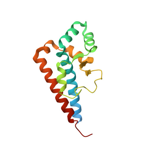
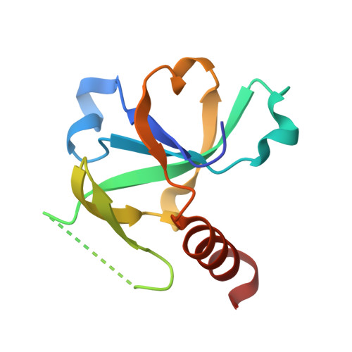
Alternative interactions define gyrase specificity in the CcdB family.

De Jonge, N.Simic, M.Buts, L.Haesaerts, S.Roelants, K.Garcia-Pino, A.Sterckx, Y.De Greve, H.Lah, J.Loris, R.

(2012) Mol Microbiol 84: 965-978

  • DOI: https://doi.org/10.1111/j.1365-2958.2012.08069.x
  • Primary Citation Related Structures: 
    4ELY, 4ELZ

  • PubMed Abstract: 

    Toxin-antitoxin (TA) modules are small operons associated with stress response of bacteria. F-plasmid CcdB(F) was the first TA toxin for which its target, gyrase, was identified. Plasmidic and chromosomal CcdBs belong to distinct families. Conserved residues crucial for gyrase poisoning activity of plasmidic CcdBs are not conserved among these families. Here we show that the chromosomal CcdB(Vfi) from Vibrio fischeri is an active gyrase poison that interacts with its target via an alternative energetic mechanism. Changes in the GyrA14-binding surface of the Vibrio and F-plasmid CcdB family members illustrate neutral drift where alternative interactions can be used to achieve the same functionality. Differences in affinity between V. fischeri and F-plasmid CcdB for gyrase and their corresponding CcdA antitoxin possibly reflect distinct roles for TA modules located on plasmids and chromosomes.


  • Organizational Affiliation
    • Structural Biology Brussels, Department of Biotechnology, Vrije Universiteit Brussel, Pleinlaan 2, B-1050 Brussels, Belgium.

Macromolecule Content 

  • Total Structure Weight: 59.06 kDa 
  • Atom Count: 3,906 
  • Modeled Residue Count: 466 
  • Deposited Residue Count: 522 
  • Unique protein chains: 2

Macromolecules

Find similar proteins by:|  3D Structure
Entity ID: 1
MoleculeChains  Sequence LengthOrganismDetailsImage
DNA gyrase subunit A
A, B
156Shigella flexneriMutation(s): 0 
Gene Names: gyrAGYRASES2444SF2311
EC: 5.99.1.3 (PDB Primary Data), 5.6.2.2 (UniProt)
UniProt
Find proteins for P0AES5 (Shigella flexneri)
Explore P0AES5 
Go to UniProtKB:  P0AES5
Entity Groups
Sequence Clusters30% Identity50% Identity70% Identity90% Identity95% Identity100% Identity
UniProt GroupP0AES5
Sequence Annotations
Expand
Reference Sequence
Find similar proteins by:|  3D Structure
Entity ID: 2
MoleculeChains  Sequence LengthOrganismDetailsImage
CcdB
C, D
105Aliivibrio fischeriMutation(s): 0 
Gene Names: ccdB
UniProt
Find proteins for Q84B82 (Aliivibrio fischeri)
Explore Q84B82 
Go to UniProtKB:  Q84B82
Entity Groups
Sequence Clusters30% Identity50% Identity70% Identity90% Identity95% Identity100% Identity
UniProt GroupQ84B82
Sequence Annotations
Expand
Reference Sequence

Experimental Data & Validation

Experimental Data

  • Method: X-RAY DIFFRACTION
  • Resolution: 1.93 Å
  • R-Value Free:  0.217 (Depositor), 0.217 (DCC) 
  • R-Value Work:  0.185 (Depositor), 0.181 (DCC) 
  • R-Value Observed: 0.187 (Depositor) 
Space Group: C 1 2 1
Unit Cell:
Length ( Å )Angle ( ˚ )
a = 130.073α = 90
b = 90.833β = 102.59
c = 58.13γ = 90
Software Package:
Software NamePurpose
ADSCdata collection
PHASERphasing
PHENIXrefinement
DENZOdata reduction
SCALEPACKdata scaling

Structure Validation

View Full Validation Report


Entry History 

Deposition Data

Revision History  (Full details and data files)

  • Version 1.0: 2012-05-30
    Type: Initial release
  • Version 1.1: 2013-01-02
    Changes: Database references
  • Version 1.2: 2023-09-13
    Changes: Data collection, Database references, Derived calculations, Refinement description