4ELC | pdb_00004elc

Crystal structure of the catalytic domain of botulinum neurotoxin BoNT/A C134 mutant with MTSEA modified Cys-165


Experimental Data Snapshot

  • Method: X-RAY DIFFRACTION
  • Resolution: 1.80 Å
  • R-Value Free: 
    0.223 (Depositor), 0.224 (DCC) 
  • R-Value Work: 
    0.169 (Depositor), 0.173 (DCC) 
  • R-Value Observed: 
    0.172 (Depositor) 

Starting Model: experimental
View more details

wwPDB Validation 3D Report Full Report

Validation slider image for 4ELC

This is version 1.4 of the entry. See complete history

Literature

Structural Framework for Covalent Inhibition of Clostridium botulinum Neurotoxin A by Targeting Cys165.

Stura, E.A.Le Roux, L.Guitot, K.Garcia, S.Bregant, S.Beau, F.Vera, L.Collet, G.Ptchelkine, D.Bakirci, H.Dive, V.

(2012) J Biological Chem 287: 33607-33614

  • DOI: https://doi.org/10.1074/jbc.M112.396697
  • Primary Citation Related Structures: 
    4EJ5, 4EL4, 4ELC

  • PubMed Abstract: 

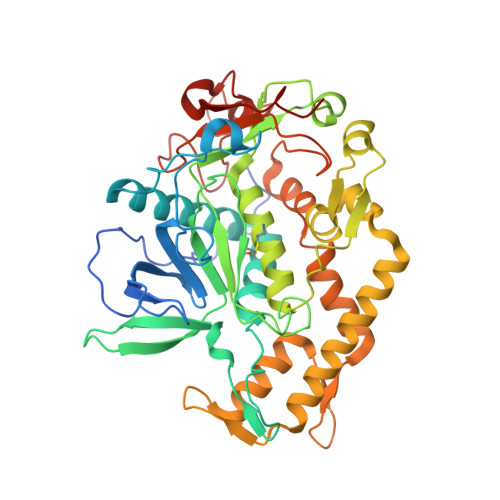
    Clostridium botulinum neurotoxin type A (BoNT/A) is one of the most potent toxins for humans and a major biothreat agent. Despite intense chemical efforts over the past 10 years to develop inhibitors of its catalytic domain (catBoNT/A), highly potent and selective inhibitors are still lacking. Recently, small inhibitors were reported to covalently modify catBoNT/A by targeting Cys(165), a residue located in the enzyme active site just above the catalytic zinc ion. However, no direct proof of Cys(165) modification was reported, and the poor accessibility of this residue in the x-ray structure of catBoNT/A raises concerns about this proposal. To clarify this issue, the functional role of Cys(165) was first assessed through a combination of site-directed mutagenesis and structural studies. These data suggested that Cys(165) is more involved in enzyme catalysis rather than in structural property. Then by peptide mass fingerprinting and x-ray crystallography, we demonstrated that a small compound containing a sulfonyl group acts as inhibitor of catBoNT/A through covalent modification of Cys(165). The crystal structure of this covalent complex offers a structural framework for developing more potent covalent inhibitors catBoNT/A. Other zinc metalloproteases can be founded in the protein database with a cysteine at a similar location, some expressed by major human pathogens; thus this work should find broader applications for developing covalent inhibitors.


  • Organizational Affiliation
    • Commissariat à l'Energie Atomique, iBiTec-S, Service d'Ingénierie Moléculaire des Protéines, CE-Saclay, 91191 Gif-sur-Yvette, Cedex, France.

Macromolecule Content 

  • Total Structure Weight: 51.99 kDa 
  • Atom Count: 4,163 
  • Modeled Residue Count: 433 
  • Deposited Residue Count: 445 
  • Unique protein chains: 1

Macromolecules

Find similar proteins by:|  3D Structure
Entity ID: 1
MoleculeChains  Sequence LengthOrganismDetailsImage
Botulinum neurotoxin A light chain445Clostridium botulinumMutation(s): 2 
Gene Names: atxbnabont/abotA
EC: 3.4.24.69
UniProt
Find proteins for P0DPI1 (Clostridium botulinum (strain Hall / ATCC 3502 / NCTC 13319 / Type A))
Explore P0DPI1 
Go to UniProtKB:  P0DPI1
Entity Groups
Sequence Clusters30% Identity50% Identity70% Identity90% Identity95% Identity100% Identity
UniProt GroupP0DPI1
Sequence Annotations
Expand
Reference Sequence

Small Molecules

Ligands 7 Unique
IDChains Name / Formula / InChI Key2D Diagram3D Interactions
LMR

Query on LMR


Download:Ideal Coordinates CCD File
E [auth A](2S)-2-hydroxybutanedioic acid
C4 H6 O5
BJEPYKJPYRNKOW-REOHCLBHSA-N
MPD

Query on MPD


Download:Ideal Coordinates CCD File
I [auth A],
J [auth A]
(4S)-2-METHYL-2,4-PENTANEDIOL
C6 H14 O2
SVTBMSDMJJWYQN-YFKPBYRVSA-N
SO4

Query on SO4


Download:Ideal Coordinates CCD File
G [auth A],
H [auth A]
SULFATE ION
O4 S
QAOWNCQODCNURD-UHFFFAOYSA-L
DMS

Query on DMS


Download:Ideal Coordinates CCD File
F [auth A],
K [auth A],
L [auth A]
DIMETHYL SULFOXIDE
C2 H6 O S
IAZDPXIOMUYVGZ-UHFFFAOYSA-N
IMD

Query on IMD


Download:Ideal Coordinates CCD File
C [auth A],
D [auth A]
IMIDAZOLE
C3 H5 N2
RAXXELZNTBOGNW-UHFFFAOYSA-O
ZN

Query on ZN


Download:Ideal Coordinates CCD File
B [auth A]ZINC ION
Zn
PTFCDOFLOPIGGS-UHFFFAOYSA-N
LI

Query on LI


Download:Ideal Coordinates CCD File
M [auth A],
N [auth A],
O [auth A]
LITHIUM ION
Li
HBBGRARXTFLTSG-UHFFFAOYSA-N
Modified Residues  1 Unique
IDChains TypeFormula2D DiagramParent
0QL
Query on 0QL
A
L-PEPTIDE LINKINGC5 H12 N2 O2 S2CYS

Experimental Data & Validation

Experimental Data

  • Method: X-RAY DIFFRACTION
  • Resolution: 1.80 Å
  • R-Value Free:  0.223 (Depositor), 0.224 (DCC) 
  • R-Value Work:  0.169 (Depositor), 0.173 (DCC) 
  • R-Value Observed: 0.172 (Depositor) 
Space Group: P 43 21 2
Unit Cell:
Length ( Å )Angle ( ˚ )
a = 65.29α = 90
b = 65.29β = 90
c = 200.91γ = 90
Software Package:
Software NamePurpose
DNAdata collection
MOLREPphasing
REFMACrefinement
XDSdata reduction
XSCALEdata scaling

Structure Validation

View Full Validation Report


Entry History 

Deposition Data

Revision History  (Full details and data files)

  • Version 1.0: 2012-08-15
    Type: Initial release
  • Version 1.1: 2012-08-29
    Changes: Database references
  • Version 1.2: 2012-10-17
    Changes: Database references
  • Version 1.3: 2022-12-21
    Changes: Database references, Derived calculations
  • Version 1.4: 2023-09-20
    Changes: Data collection, Refinement description