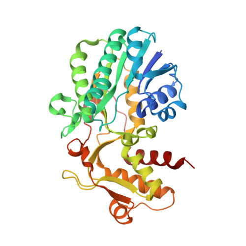
Structure and in silico substrate-binding mode of ADP-L-glycero-D-manno-heptose 6-epimerase from Burkholderia thailandensis.
Kim, M.S., Lim, A., Yang, S.W., Park, J., Lee, D., Shin, D.H.(2013) Acta Crystallogr D Biol Crystallogr 69: 658-668
- PubMed: 23519675 Search on PubMed
- DOI: https://doi.org/10.1107/S0907444913001030
- Primary Citation Related Structures:
4EJ0 - PubMed Abstract:
ADP-L-glycero-D-manno-heptose 6-epimerase (AGME), the product of the rfaD gene, is the last enzyme in the heptose-biosynthesis pathway; it converts ADP-D-glycero-D-manno-heptose (ADP-D,D-Hep) to ADP-L-glycero-D-manno-heptose (ADP-L,D-Hep). AGME contains a catalytic triad involved in catalyzing hydride transfer with the aid of NADP(+). Defective lipopolysaccharide is found in bacterial mutants lacking this gene. Therefore, it is an interesting target enzyme for a novel epimerase inhibitor for use as a co-therapy with antibiotics. The crystal structure of AGME from Burkholderia thailandensis (BtAGME), a surrogate organism for studying the pathogenicity of melioidosis caused by B. pseudomallei, has been determined. The crystal structure determined with co-purified NADP(+) revealed common as well as unique structural properties of the AGME family when compared with UDP-galactose 4-epimerase homologues. They form a similar architecture with conserved catalytic residues. Nevertheless, there are differences in the substrate- and cofactor-binding cavities and the oligomerization domains. Structural comparison of BtAGME with AGME from Escherichia coli indicates that they may recognize their substrate in a `lock-and-key' fashion. Unique structural features of BtAGME are found in two regions. The first region is the loop between β8 and β9, affecting the binding affinity of BtAGME for the ADP moiety of ADP-D,D-Hep. The second region is helix α8, which induces decamerization at low pH that is not found in other AGMEs. With the E210G mutant, it was observed that the resistance of the wild type to acid-induced denaturation is related to the decameric state. An in silico study was performed using the Surflex-Dock GeomX module of the SYBYL-X 1.3 software to predict the catalytic mechanism of BtAGME with its substrate, ADP-D,D-Hep. In the in silico study, the C7'' hydroxymethyl group of ADP-D,D-Hep is predicted to form hydrogen bonds to Ser116 and Gln293. With the aid of these interactions, the hydroxyl of Tyr139 forms a hydrogen bond to O6″ of ADP-D,D-Hep and the proton at C6″ orients closely to C4 of NADP(+). Therefore, the in silico study supports a one-base mechanism as a major catalytic pathway, in which Tyr139 solely functions as a catalytic acid/base residue. These results provide a new insight into the development of an epimerase inhibitor as an antibiotic adjuvant against melioidosis.
- The Center for Cell Signaling and Drug Discovery Research, College of Pharmacy, Division of Life and Pharmaceutical Sciences, Ewha Womans University, Seoul, Republic of Korea.
Organizational Affiliation: