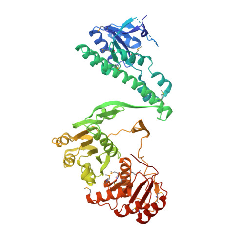
An X-ray Crystal Structure of a putative Bifunctional Phosphoribosylaminoimidazolecarboxamide Formyltransferase/IMP Cyclohydrolase
Brunzelle, J.S., Wawrzak, Z., Onopriyenko, O., Kwok, J., Anderson, W.F., Savchenko, A., Center for Structural Genomics of Infectious DiseasesTo be published.