


2.50 angstrom resolution structure of 3-phosphoshikimate 1-carboxyvinyltransferase (AroA) from Coxiella burnetii in complex with phosphoenolpyruvate
Krishna, S.N., Light, S.H., Minasov, G., Shuvalova, L., Kwon, K., Anderson, W.F.To be published.
Experimental Data Snapshot
Starting Model: experimental
View more details
Macromolecule Content
Entity ID: 1 | |||||
|---|---|---|---|---|---|
| Molecule | Chains | Sequence Length | Organism | Details | Image |
| 3-phosphoshikimate 1-carboxyvinyltransferase | 441 | Coxiella burnetii | Mutation(s): 0 Gene Names: aroA, CBU_0526 EC: 2.5.1.19 | 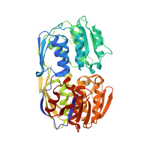 | |
UniProt | |||||
Entity Groups | |||||
| Sequence Clusters | 30% Identity50% Identity70% Identity90% Identity95% Identity100% Identity | ||||
| UniProt Group | Q83E11 | ||||
Sequence AnnotationsExpand | |||||
Reference Sequence | |||||
| Ligands 2 Unique | |||||
|---|---|---|---|---|---|
| ID | Chains | Name / Formula / InChI Key | 2D Diagram | 3D Interactions | |
| PEP Download:Ideal Coordinates CCD File | G [auth A] L [auth B] O [auth C] R [auth D] U [auth E] | PHOSPHOENOLPYRUVATE C3 H5 O6 P DTBNBXWJWCWCIK-UHFFFAOYSA-N |  | ||
| SO4 Download:Ideal Coordinates CCD File | H [auth A] I [auth A] J [auth B] K [auth B] M [auth C] | SULFATE ION O4 S QAOWNCQODCNURD-UHFFFAOYSA-L |  | ||
| Length ( Å ) | Angle ( ˚ ) |
|---|---|
| a = 162.363 | α = 90 |
| b = 94.05 | β = 90.01 |
| c = 230.51 | γ = 90 |
| Software Name | Purpose |
|---|---|
| HKL-2000 | data collection |
| PHASER | phasing |
| REFMAC | refinement |
| HKL-3000 | data reduction |
| HKL-3000 | data scaling |


