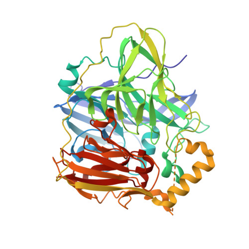
4EF3

Multicopper Oxidase CueO (Citrate buffer)


Experimental Data Snapshot

  • Method: X-RAY DIFFRACTION
  • Resolution: 1.90 Å
  • R-Value Free: 0.184 
  • R-Value Work: 0.152 
  • R-Value Observed: 0.154 

wwPDB Validation   3D Report Full Report


This is version 1.3 of the entry. See complete history


Literature

Exogenous acetate ion reaches the type II copper centre in CueO through the water-excretion channel and potentially affects the enzymatic activity.

Komori, H.Kataoka, K.Tanaka, S.Matsuda, N.Higuchi, Y.Sakurai, T.

(2016) Acta Crystallogr Sect F Struct Biol Cryst Commun 72: 558-563

  • DOI: https://doi.org/10.1107/S2053230X16009237
  • Primary Citation of Related Structures:  
    4EF3

  • PubMed Abstract: 

    The acetate-bound form of the type II copper was found in the X-ray structure of the multicopper oxidase CueO crystallized in acetate buffer in addition to the conventional OH(-)-bound form as the major resting form. The acetate ion was retained bound to the type II copper even after prolonged exposure of a CueO crystal to X-ray radiation, which led to the stepwise reduction of the Cu centres. However, in this study, when CueO was crystallized in citrate buffer the OH(-)-bound form was present exclusively. This fact shows that an exogenous acetate ion reaches the type II Cu centre through the water channel constructed between domains 1 and 3 in the CueO molecule. It was also found that the enzymatic activity of CueO is enhanced in the presence of acetate ions in the solvent water.


  • Organizational Affiliation

    Faculty of Education, Kagawa University, 1-1 Saiwai-cho, Takamatsu, Kagawa 760-8522, Japan.


Macromolecules
Find similar proteins by:  (by identity cutoff)  |  3D Structure
Entity ID: 1
MoleculeChains Sequence LengthOrganismDetailsImage
Blue copper oxidase CueO489Escherichia coli K-12Mutation(s): 0 
Gene Names: cueOyacKb0123JW0119
EC: 1.16.3.4
UniProt
Find proteins for P36649 (Escherichia coli (strain K12))
Explore P36649 
Go to UniProtKB:  P36649
Entity Groups  
Sequence Clusters30% Identity50% Identity70% Identity90% Identity95% Identity100% Identity
UniProt GroupP36649
Sequence Annotations
Expand
  • Reference Sequence
Small Molecules
Ligands 4 Unique
IDChains Name / Formula / InChI Key2D Diagram3D Interactions
CIT
Query on CIT

Download Ideal Coordinates CCD File 
H [auth A]CITRIC ACID
C6 H8 O7
KRKNYBCHXYNGOX-UHFFFAOYSA-N
CU
Query on CU

Download Ideal Coordinates CCD File 
B [auth A],
C [auth A],
D [auth A],
E [auth A]
COPPER (II) ION
Cu
JPVYNHNXODAKFH-UHFFFAOYSA-N
OH
Query on OH

Download Ideal Coordinates CCD File 
F [auth A]HYDROXIDE ION
H O
XLYOFNOQVPJJNP-UHFFFAOYSA-M
O
Query on O

Download Ideal Coordinates CCD File 
G [auth A]OXYGEN ATOM
O
XLYOFNOQVPJJNP-UHFFFAOYSA-N
Experimental Data & Validation

Experimental Data

  • Method: X-RAY DIFFRACTION
  • Resolution: 1.90 Å
  • R-Value Free: 0.184 
  • R-Value Work: 0.152 
  • R-Value Observed: 0.154 
  • Space Group: P 1 21 1
Unit Cell:
Length ( Å )Angle ( ˚ )
a = 50.282α = 90
b = 91.239β = 112.87
c = 59.589γ = 90
Software Package:
Software NamePurpose
REFMACrefinement

Structure Validation

View Full Validation Report


Entry History 

Deposition Data

Revision History  (Full details and data files)

  • Version 1.0: 2013-05-22
    Type: Initial release
  • Version 1.1: 2013-07-24
    Changes: Database references
  • Version 1.2: 2016-08-03
    Changes: Database references
  • Version 1.3: 2024-03-20
    Changes: Data collection, Database references, Derived calculations