4E9C | pdb_00004e9c

The structure of the polo-box domain (PBD) of polo-like kinase 1 (Plk1) in complex with LDPPLHSpTA phosphopeptide


Experimental Data Snapshot

  • Method: X-RAY DIFFRACTION
  • Resolution: 1.70 Å
  • R-Value Free: 
    0.232 (Depositor), 0.232 (DCC) 
  • R-Value Work: 
    0.192 (Depositor) 
  • R-Value Observed: 
    0.194 (Depositor) 

Starting Model: experimental
View more details

wwPDB Validation 3D Report Full Report

Validation slider image for 4E9C

This is version 1.5 of the entry. See complete history

Literature

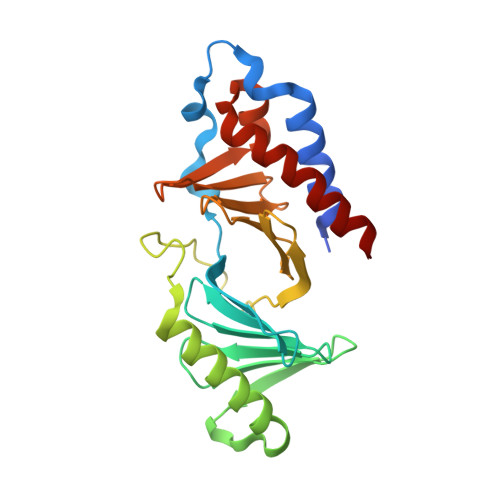
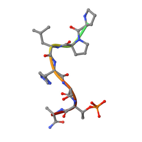
High-throughput interrogation of ligand binding mode using a fluorescence-based assay.

Sledz, P.Lang, S.Stubbs, C.J.Abell, C.

(2012) Angew Chem Int Ed Engl 51: 7680-7683

  • DOI: https://doi.org/10.1002/anie.201202660
  • Primary Citation Related Structures: 
    4E9C, 4E9D

  • PubMed Abstract: 

    Probing the pocket: A high-throughput fluorescence-based thermal shift (FTS) assay utilized different forms of a protein (in gray) to establish the binding mode of a ligand (see picture). The assay serves in the rapid evaluation of structure-activity binding-mode relationships for a series of ligands of Plk1, an important target of anticancer therapy.


  • Organizational Affiliation
    • University Chemical Laboratory, University of Cambridge, Lensfield Road, Cambridge CB2 1EW, UK.

Macromolecule Content 

  • Total Structure Weight: 28.14 kDa 
  • Atom Count: 2,076 
  • Modeled Residue Count: 231 
  • Deposited Residue Count: 243 
  • Unique protein chains: 2

Macromolecules

Find similar proteins by:|  3D Structure
Entity ID: 1
MoleculeChains  Sequence LengthOrganismDetailsImage
Serine/threonine-protein kinase PLK1232Homo sapiensMutation(s): 0 
Gene Names: PLK1PLK
EC: 2.7.11.21
UniProt & NIH Common Fund Data Resources
Find proteins for P53350 (Homo sapiens)
Explore P53350 
Go to UniProtKB:  P53350
PHAROS:  P53350
GTEx:  ENSG00000166851 
Entity Groups
Sequence Clusters30% Identity50% Identity70% Identity90% Identity95% Identity100% Identity
UniProt GroupP53350
Sequence Annotations
Expand
Reference Sequence
Find similar proteins by:  Sequence   |   3D Structure  
Entity ID: 2
MoleculeChains  Sequence LengthOrganismDetailsImage
LDPPLHSpTA phosphopeptide11N/AMutation(s): 0 

Small Molecules

Biologically Interesting Molecules (External Reference) 

1 Unique

Experimental Data & Validation

Experimental Data

  • Method: X-RAY DIFFRACTION
  • Resolution: 1.70 Å
  • R-Value Free:  0.232 (Depositor), 0.232 (DCC) 
  • R-Value Work:  0.192 (Depositor) 
  • R-Value Observed: 0.194 (Depositor) 
Space Group: P 1 21 1
Unit Cell:
Length ( Å )Angle ( ˚ )
a = 35.634α = 90
b = 89.784β = 109.28
c = 37.108γ = 90
Software Package:
Software NamePurpose
MOLREPphasing
REFMACrefinement
XDSdata reduction
XDSdata scaling

Structure Validation

View Full Validation Report


Entry History 

Deposition Data

Revision History  (Full details and data files)

  • Version 1.0: 2012-10-10
    Type: Initial release
  • Version 1.1: 2012-10-17
    Changes: Database references
  • Version 1.2: 2012-12-12
    Changes: Other
  • Version 1.3: 2018-12-05
    Changes: Data collection, Database references
  • Version 1.4: 2023-09-13
    Changes: Data collection, Database references, Derived calculations, Refinement description
  • Version 1.5: 2024-11-06
    Changes: Structure summary