4E80 | pdb_00004e80

Structural Basis for the Activity of a Cytoplasmic RNA Terminal U-transferase


Experimental Data Snapshot

  • Method: X-RAY DIFFRACTION
  • Resolution: 3.02 Å
  • R-Value Free: 
    0.201 (Depositor), 0.204 (DCC) 
  • R-Value Work: 
    0.176 (Depositor), 0.182 (DCC) 
  • R-Value Observed: 
    0.178 (Depositor) 

wwPDB Validation 3D Report Full Report

Validation slider image for 4E80

Ligand Structure Quality Assessment 


This is version 1.4 of the entry. See complete history

Literature

Structural basis for the activity of a cytoplasmic RNA terminal uridylyl transferase.

Yates, L.A.Fleurdepine, S.Rissland, O.S.De Colibus, L.Harlos, K.Norbury, C.J.Gilbert, R.J.

(2012) Nat Struct Mol Biol 19: 782-787

  • DOI: https://doi.org/10.1038/nsmb.2329
  • Primary Citation Related Structures: 
    4E7X, 4E80, 4E8F

  • PubMed Abstract: 

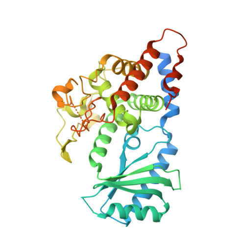
    Cytoplasmic terminal uridylyl transferases comprise a conserved family of enzymes that negatively regulate the stability or biological activity of a variety of eukaryotic RNAs, including mRNAs and tumor-suppressor let-7 microRNAs. Here we describe crystal structures of the Schizosaccharomyces pombe cytoplasmic terminal uridylyl transferase Cid1 in two apo conformers and bound to UTP. We demonstrate that a single histidine residue, conserved in mammalian Cid1 orthologs, is responsible for discrimination between UTP and ATP. We also describe a new high-affinity RNA substrate-binding mechanism of Cid1, which is essential for enzymatic activity and is mediated by three basic patches across the surface of the enzyme. Overall, our structures provide a basis for understanding the activity of Cid1 and a mechanism of UTP selectivity conserved in its human orthologs, suggesting potential implications for anticancer drug design.


  • Organizational Affiliation
    • Division of Structural Biology, Wellcome Trust Centre for Human Genetics, University of Oxford, Roosevelt Drive, Oxford OX3 7BN, UK.

Macromolecule Content 

  • Total Structure Weight: 187.22 kDa 
  • Atom Count: 10,472 
  • Modeled Residue Count: 1,282 
  • Deposited Residue Count: 1,620 
  • Unique protein chains: 1

Macromolecules

Find similar proteins by:|  3D Structure
Entity ID: 1
MoleculeChains  Sequence LengthOrganismDetailsImage
Poly(A) RNA polymerase protein cid1
A, B, C, D
405Schizosaccharomyces pombe 972h-Mutation(s): 0 
Gene Names: cid1SPAC19D5.03
EC: 2.7.7 (PDB Primary Data), 2.7.7.19 (UniProt), 2.7.7.52 (UniProt)
UniProt
Find proteins for O13833 (Schizosaccharomyces pombe (strain 972 / ATCC 24843))
Explore O13833 
Go to UniProtKB:  O13833
Entity Groups
Sequence Clusters30% Identity50% Identity70% Identity90% Identity95% Identity100% Identity
UniProt GroupO13833
Sequence Annotations
Expand
Reference Sequence

Experimental Data & Validation

Experimental Data

  • Method: X-RAY DIFFRACTION
  • Resolution: 3.02 Å
  • R-Value Free:  0.201 (Depositor), 0.204 (DCC) 
  • R-Value Work:  0.176 (Depositor), 0.182 (DCC) 
  • R-Value Observed: 0.178 (Depositor) 
Space Group: C 1 2 1
Unit Cell:
Length ( Å )Angle ( ˚ )
a = 164.69α = 90
b = 77.96β = 109.29
c = 151.68γ = 90
Software Package:
Software NamePurpose
PHASERphasing
BUSTERrefinement
XDSdata reduction
SCALAdata scaling

Structure Validation

View Full Validation Report


Ligand Structure Quality Assessment 


Entry History 

Deposition Data

Revision History  (Full details and data files)

  • Version 1.0: 2012-07-04
    Type: Initial release
  • Version 1.1: 2012-07-18
    Changes: Database references
  • Version 1.2: 2012-08-22
    Changes: Database references
  • Version 1.3: 2017-11-15
    Changes: Refinement description
  • Version 1.4: 2024-02-28
    Changes: Data collection, Database references, Derived calculations