4E6N

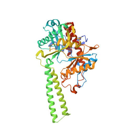
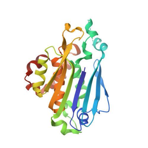
Crystal structure of bacterial Pnkp-C/Hen1-N heterodimer


Experimental Data Snapshot

  • Method: X-RAY DIFFRACTION
  • Resolution: 2.39 Å
  • R-Value Free: 0.223 
  • R-Value Work: 0.168 
  • R-Value Observed: 0.170 

wwPDB Validation   3D Report Full Report


Ligand Structure Quality Assessment 


This is version 1.3 of the entry. See complete history


Literature

Molecular basis of bacterial protein Hen1 activating the ligase activity of bacterial protein Pnkp for RNA repair.

Wang, P.Chan, C.M.Christensen, D.Zhang, C.Selvadurai, K.Huang, R.H.

(2012) Proc Natl Acad Sci U S A 109: 13248-13253

  • DOI: https://doi.org/10.1073/pnas.1209805109
  • Primary Citation of Related Structures:  
    4DQZ, 4DRF, 4E6N

  • PubMed Abstract: 

    Ribotoxins cleave essential RNAs for cell killing in vivo, and the bacterial polynucleotide kinase-phosphatase (Pnkp)/hua enhancer 1 (Hen1) complex has been shown to repair ribotoxin-cleaved RNAs in vitro. Bacterial Pnkp/Hen1 is distinguished from other RNA repair systems by performing 3'-terminal 2'-O-methylation during RNA repair, which prevents the repaired RNA from repeated cleavage at the same site. To ensure the opportunity of 2'-O-methylation by bacterial Hen1 during RNA repair and, therefore, maintain the quality of the repaired RNA, Pnkp/Hen1 has evolved to require the participation of Hen1 in RNA ligation, because Pnkp alone is unable to carry out the reaction despite possessing all signature motifs of an RNA ligase. However, the precise role of Hen1 in RNA ligation is unknown. Here, we present the crystal structure of an active RNA ligase consisting of the C-terminal half of Pnkp (Pnkp-C) and the N-terminal half of Hen1 (Hen1-N) from Clostridium thermocellum. The structure reveals that the N-terminal domain of Clostridium thermocellum (Cth) Hen1, shaped like a left hand, grabs the flexible insertion module of CthPnkp and locks its conformation via further interaction with the C-terminal addition module of CthPnkp. Formation of the CthPnkp-C/Hen1-N heterodimer creates a ligation pocket with a width for two strands of RNA, depth for two nucleotides, and the adenosine monophosphate (AMP)-binding pocket at the bottom. The structure, combined with functional analyses, provides insight into the mechanism of how Hen1 activates the RNA ligase activity of Pnkp for RNA repair.


  • Organizational Affiliation

    Department of Biochemistry, University of Illinois at Urbana-Champaign, Urbana, IL 61801, USA.


Macromolecules
Find similar proteins by:  (by identity cutoff)  |  3D Structure
Entity ID: 1
MoleculeChains Sequence LengthOrganismDetailsImage
Metallophosphoesterase
A, C
427Acetivibrio thermocellus ATCC 27405Mutation(s): 0 
Gene Names: Cthe_2768
UniProt
Find proteins for A3DJ38 (Acetivibrio thermocellus (strain ATCC 27405 / DSM 1237 / JCM 9322 / NBRC 103400 / NCIMB 10682 / NRRL B-4536 / VPI 7372))
Explore A3DJ38 
Go to UniProtKB:  A3DJ38
Entity Groups  
Sequence Clusters30% Identity50% Identity70% Identity90% Identity95% Identity100% Identity
UniProt GroupA3DJ38
Sequence Annotations
Expand
  • Reference Sequence
Find similar proteins by:  (by identity cutoff)  |  3D Structure
Entity ID: 2
MoleculeChains Sequence LengthOrganismDetailsImage
Methyltransferase type 12
B, D
230Acetivibrio thermocellus ATCC 27405Mutation(s): 0 
Gene Names: Cthe_2767
EC: 2.1.1.386
UniProt
Find proteins for A3DJ37 (Acetivibrio thermocellus (strain ATCC 27405 / DSM 1237 / JCM 9322 / NBRC 103400 / NCIMB 10682 / NRRL B-4536 / VPI 7372))
Explore A3DJ37 
Go to UniProtKB:  A3DJ37
Entity Groups  
Sequence Clusters30% Identity50% Identity70% Identity90% Identity95% Identity100% Identity
UniProt GroupA3DJ37
Sequence Annotations
Expand
  • Reference Sequence
Experimental Data & Validation

Experimental Data

  • Method: X-RAY DIFFRACTION
  • Resolution: 2.39 Å
  • R-Value Free: 0.223 
  • R-Value Work: 0.168 
  • R-Value Observed: 0.170 
  • Space Group: P 1
Unit Cell:
Length ( Å )Angle ( ˚ )
a = 59.63α = 81.38
b = 59.695β = 87.82
c = 101.54γ = 88.82
Software Package:
Software NamePurpose
ADSCdata collection
PHENIXmodel building
PHENIXrefinement
HKL-2000data reduction
HKL-2000data scaling
PHENIXphasing

Structure Validation

View Full Validation Report


Ligand Structure Quality Assessment 


Entry History 

Deposition Data

Revision History  (Full details and data files)

  • Version 1.0: 2012-08-01
    Type: Initial release
  • Version 1.1: 2012-08-15
    Changes: Database references
  • Version 1.2: 2012-08-29
    Changes: Database references
  • Version 1.3: 2024-02-28
    Changes: Data collection, Database references, Derived calculations