4DYD | pdb_00004dyd

Substrate-directed dual catalysis of dicarbonyl compounds by diketoreductase


Experimental Data Snapshot

  • Method: X-RAY DIFFRACTION
  • Resolution: 1.95 Å
  • R-Value Free: 
    0.242 (Depositor), 0.243 (DCC) 
  • R-Value Work: 
    0.217 (Depositor), 0.217 (DCC) 
  • R-Value Observed: 
    0.217 (Depositor) 

wwPDB Validation 3D Report Full Report

Validation slider image for 4DYD

Ligand Structure Quality Assessment 


This is version 1.1 of the entry. See complete history

Literature

Dual catalysis mode for the dicarbonyl reduction catalyzed by diketoreductase

Lu, M.Huang, Y.White, M.A.Wu, X.Liu, N.Cheng, X.Chen, Y.

(2012) Chem Commun (Camb) 48: 11352-11354

  • DOI: https://doi.org/10.1039/c2cc36334h
  • Primary Citation Related Structures: 
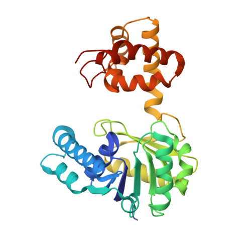
    4DYD, 4E12, 4E13

  • PubMed Abstract: 

    Diketoreductase catalyzes a two-step bioreduction on a dicarbonyl substrate through a novel dual catalysis mode, in which random hydride attack simultaneously forms two mono-carbonyl intermediates, and subsequently distinct catalytic sites are responsible for the reductions of respective carbonyl group of the intermediates to yield the final dihydroxy product.


  • Organizational Affiliation
    • Laboratory of Chemical Biology, China Pharmaceutical University, 24 Tongjia Street, Nanjing, Jiangsu Province 210009, People's Republic of China.

Macromolecule Content 

  • Total Structure Weight: 32.14 kDa 
  • Atom Count: 2,296 
  • Modeled Residue Count: 283 
  • Deposited Residue Count: 283 
  • Unique protein chains: 1

Macromolecules

Find similar proteins by:|  3D Structure
Entity ID: 1
MoleculeChains  Sequence LengthOrganismDetailsImage
Diketoreductase283Acinetobacter baylyiMutation(s): 0 
UniProt
Find proteins for B1P3E1 (Acinetobacter baylyi)
Explore B1P3E1 
Go to UniProtKB:  B1P3E1
Entity Groups
Sequence Clusters30% Identity50% Identity70% Identity90% Identity95% Identity100% Identity
UniProt GroupB1P3E1
Sequence Annotations
Expand
Reference Sequence

Small Molecules

Ligands 4 Unique
IDChains Name / Formula / InChI Key2D Diagram3D Interactions
NAD

Query on NAD


Download:Ideal Coordinates CCD File
B [auth A]NICOTINAMIDE-ADENINE-DINUCLEOTIDE
C21 H27 N7 O14 P2
BAWFJGJZGIEFAR-NNYOXOHSSA-N
SO4

Query on SO4


Download:Ideal Coordinates CCD File
M [auth A]SULFATE ION
O4 S
QAOWNCQODCNURD-UHFFFAOYSA-L
PO4

Query on PO4


Download:Ideal Coordinates CCD File
N [auth A],
O [auth A]
PHOSPHATE ION
O4 P
NBIIXXVUZAFLBC-UHFFFAOYSA-K
GOL

Query on GOL


Download:Ideal Coordinates CCD File
C [auth A]
D [auth A]
E [auth A]
F [auth A]
G [auth A]
C [auth A],
D [auth A],
E [auth A],
F [auth A],
G [auth A],
H [auth A],
I [auth A],
J [auth A],
K [auth A],
L [auth A]
GLYCEROL
C3 H8 O3
PEDCQBHIVMGVHV-UHFFFAOYSA-N

Experimental Data & Validation

Experimental Data

  • Method: X-RAY DIFFRACTION
  • Resolution: 1.95 Å
  • R-Value Free:  0.242 (Depositor), 0.243 (DCC) 
  • R-Value Work:  0.217 (Depositor), 0.217 (DCC) 
  • R-Value Observed: 0.217 (Depositor) 
Space Group: P 62 2 2
Unit Cell:
Length ( Å )Angle ( ˚ )
a = 100.48α = 90
b = 100.48β = 90
c = 131.91γ = 120
Software Package:
Software NamePurpose
ADSCdata collection
SOLVEphasing
CNSrefinement
HKL-3000data reduction
HKL-3000data scaling

Structure Validation

View Full Validation Report


Ligand Structure Quality Assessment 


Entry History 

Deposition Data

Revision History  (Full details and data files)

  • Version 1.0: 2012-11-14
    Type: Initial release
  • Version 1.1: 2024-03-20
    Changes: Data collection, Database references, Derived calculations