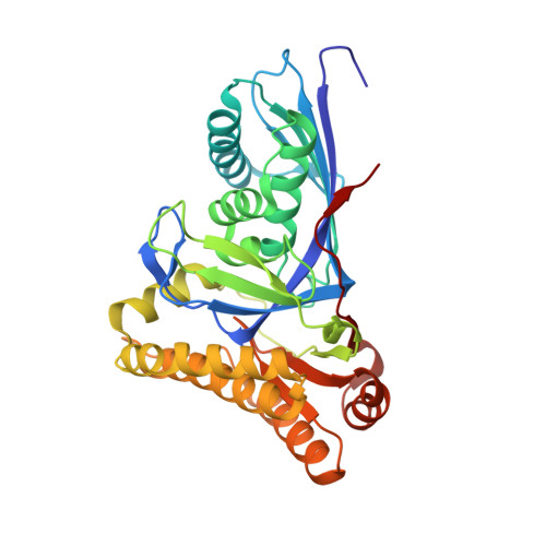
Structural basis for nucleotide binding and reaction catalysis in mevalonate diphosphate decarboxylase.
Barta, M.L., McWhorter, W.J., Miziorko, H.M., Geisbrecht, B.V.(2012) Biochemistry 51: 5611-5621
- PubMed: 22734632
- DOI: https://doi.org/10.1021/bi300591x
- Primary Citation of Related Structures:
4DPT, 4DPU, 4DPW, 4DPX, 4DPY, 4DU7, 4DU8 - PubMed Abstract:
Mevalonate diphosphate decarboxylase (MDD) catalyzes the final step of the mevalonate pathway, the Mg(2+)-ATP dependent decarboxylation of mevalonate 5-diphosphate (MVAPP), producing isopentenyl diphosphate (IPP). Synthesis of IPP, an isoprenoid precursor molecule that is a critical intermediate in peptidoglycan and polyisoprenoid biosynthesis, is essential in Gram-positive bacteria (e.g., Staphylococcus, Streptococcus, and Enterococcus spp.), and thus the enzymes of the mevalonate pathway are ideal antimicrobial targets. MDD belongs to the GHMP superfamily of metabolite kinases that have been extensively studied for the past 50 years, yet the crystallization of GHMP kinase ternary complexes has proven to be difficult. To further our understanding of the catalytic mechanism of GHMP kinases with the purpose of developing broad spectrum antimicrobial agents that target the substrate and nucleotide binding sites, we report the crystal structures of wild-type and mutant (S192A and D283A) ternary complexes of Staphylococcus epidermidis MDD. Comparison of apo, MVAPP-bound, and ternary complex wild-type MDD provides structural information about the mode of substrate binding and the catalytic mechanism. Structural characterization of ternary complexes of catalytically deficient MDD S192A and D283A (k(cat) decreased 10(3)- and 10(5)-fold, respectively) provides insight into MDD function. The carboxylate side chain of invariant Asp(283) functions as a catalytic base and is essential for the proper orientation of the MVAPP C3-hydroxyl group within the active site funnel. Several MDD amino acids within the conserved phosphate binding loop ("P-loop") provide key interactions, stabilizing the nucleotide triphosphoryl moiety. The crystal structures presented here provide a useful foundation for structure-based drug design.
- Division of Cell Biology and Biophysics, School of Biological Sciences, University of Missouri-Kansas City, Kansas City, MO 64110, USA.
Organizational Affiliation:


