4DLK | pdb_00004dlk

Crystal Structure of ATP-Ca++ complex of purK: N5-carboxyaminoimidazole ribonucleotide synthetase


Experimental Data Snapshot

  • Method: X-RAY DIFFRACTION
  • Resolution: 2.02 Å
  • R-Value Free: 
    0.235 (Depositor), 0.233 (DCC) 
  • R-Value Work: 
    0.198 (Depositor), 0.196 (DCC) 
  • R-Value Observed: 
    0.200 (Depositor) 

wwPDB Validation 3D Report Full Report

Validation slider image for 4DLK

Ligand Structure Quality Assessment 


This is version 1.2 of the entry. See complete history

Literature

Elucidation of the bicarbonate binding site and insights into the carboxylation mechanism of (N(5))-carboxyaminoimidazole ribonucleotide synthase (PurK) from Bacillus anthracis.

Tuntland, M.L.Santarsiero, B.D.Johnson, M.E.Fung, L.W.

(2014) Acta Crystallogr D Biol Crystallogr 70: 3057-3065

  • DOI: https://doi.org/10.1107/S1399004714021166
  • Primary Citation Related Structures: 
    3QFF, 3R5H, 3V4S, 4DLK

  • PubMed Abstract: 

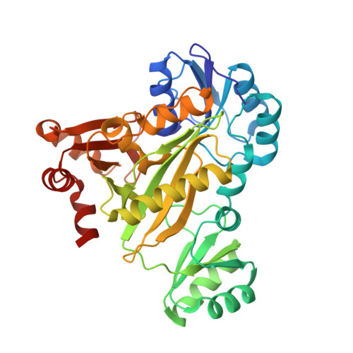
    Structures of (N(5))-carboxyaminoimidazole ribonucleotide synthase (PurK) from Bacillus anthracis with various combinations of ATP, ADP, Mg(2+), bicarbonate and aminoimidazole ribonucleotide (AIR) in the active site are presented. The binding site of bicarbonate has only been speculated upon previously, but is shown here for the first time. The binding involves interactions with the conserved residues Arg272, His274 and Lys348. These structures provide insights into each ligand in the active site and allow a possible mechanism to be proposed for the reaction that converts bicarbonate and AIR, in the presence of ATP, to produce (N(5))-carboxyaminoimidazole ribonucleotide. The formation of a carboxyphosphate intermediate through ATP phosphoryl transfer is proposed, followed by carboxylation of AIR to give the product, facilitated by a cluster of conserved residues and an active-site water network.


  • Organizational Affiliation
    • Department of Chemistry, University of Illinois at Chicago, Chicago, IL 60607, USA.

Macromolecule Content 

  • Total Structure Weight: 86.77 kDa 
  • Atom Count: 6,368 
  • Modeled Residue Count: 760 
  • Deposited Residue Count: 760 
  • Unique protein chains: 1

Macromolecules

Find similar proteins by:|  3D Structure
Entity ID: 1
MoleculeChains  Sequence LengthOrganismDetailsImage
Phosphoribosylaminoimidazole carboxylase, ATPase subunit
A, B
380Bacillus anthracis str. A0248Mutation(s): 0 
Gene Names: BAS0276BA_0289GBAA_0289purK
EC: 4.1.1.21 (PDB Primary Data), 6.3.4.18 (UniProt)
UniProt
Find proteins for A0A6L8P0X7 (Bacillus anthracis)
Explore A0A6L8P0X7 
Go to UniProtKB:  A0A6L8P0X7
Entity Groups
Sequence Clusters30% Identity50% Identity70% Identity90% Identity95% Identity100% Identity
UniProt GroupA0A6L8P0X7
Sequence Annotations
Expand
Reference Sequence

Small Molecules

Ligands 3 Unique
IDChains Name / Formula / InChI Key2D Diagram3D Interactions
ATP

Query on ATP


Download:Ideal Coordinates CCD File
F [auth A],
G [auth A],
H [auth A],
L [auth B]
ADENOSINE-5'-TRIPHOSPHATE
C10 H16 N5 O13 P3
ZKHQWZAMYRWXGA-KQYNXXCUSA-N
PO4

Query on PO4


Download:Ideal Coordinates CCD File
I [auth A]PHOSPHATE ION
O4 P
NBIIXXVUZAFLBC-UHFFFAOYSA-K
CA

Query on CA


Download:Ideal Coordinates CCD File
C [auth A],
D [auth A],
E [auth A],
J [auth B],
K [auth B]
CALCIUM ION
Ca
BHPQYMZQTOCNFJ-UHFFFAOYSA-N

Experimental Data & Validation

Experimental Data

  • Method: X-RAY DIFFRACTION
  • Resolution: 2.02 Å
  • R-Value Free:  0.235 (Depositor), 0.233 (DCC) 
  • R-Value Work:  0.198 (Depositor), 0.196 (DCC) 
  • R-Value Observed: 0.200 (Depositor) 
Space Group: P 21 21 21
Unit Cell:
Length ( Å )Angle ( ˚ )
a = 57.368α = 90
b = 84.476β = 90
c = 167.269γ = 90
Software Package:
Software NamePurpose
XSCALEdata scaling
PHASERphasing
REFMACrefinement
PDB_EXTRACTdata extraction
HKL-2000data collection

Structure Validation

View Full Validation Report


Ligand Structure Quality Assessment 


Entry History 

Deposition Data

Revision History  (Full details and data files)

  • Version 1.0: 2013-02-06
    Type: Initial release
  • Version 1.1: 2014-11-12
    Changes: Database references
  • Version 1.2: 2024-02-28
    Changes: Data collection, Database references, Derived calculations