4DFF | pdb_00004dff

The SAR development of dihydroimidazoisoquinoline derivatives as phosphodiesterase 10A inhibitors for the treatment of schizophrenia


Experimental Data Snapshot

  • Method: X-RAY DIFFRACTION
  • Resolution: 2.11 Å
  • R-Value Free: 
    0.223 (Depositor), 0.237 (DCC) 
  • R-Value Work: 
    0.190 (Depositor), 0.201 (DCC) 
  • R-Value Observed: 
    0.192 (Depositor) 

wwPDB Validation 3D Report Full Report

Validation slider image for 4DFF

Ligand Structure Quality Assessment 


This is version 1.2 of the entry. See complete history

Literature

The SAR development of dihydroimidazoisoquinoline derivatives as phosphodiesterase 10A inhibitors for the treatment of schizophrenia.

Ho, G.D.Michael Seganish, W.Bercovici, A.Tulshian, D.Greenlee, W.J.Van Rijn, R.Hruza, A.Xiao, L.Rindgen, D.Mullins, D.Guzzi, M.Zhang, X.Bleickardt, C.Hodgson, R.

(2012) Bioorg Med Chem Lett 22: 2585-2589

  • DOI: https://doi.org/10.1016/j.bmcl.2012.01.113
  • Primary Citation Related Structures: 
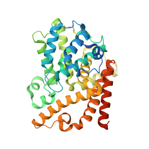
    4DFF

  • PubMed Abstract: 

    The identification of potent and orally active dihydroimidazoisoquinolines as PDE 10A inhibitors is reported. The SAR development led to the discovery of compound 35 as a potent, selective, and orally active PDE10A inhibitor. Compound 35 inhibited MK-801-induced hyperactivity at 3mg/kg and displayed a 10-fold separation between the minimal effective doses for inhibition of MK-801-induced hyperactivity and hypolocomotion in rats.


  • Organizational Affiliation
    • Department of Medicinal Chemistry, Merck Research Laboratories, Kenilworth, NJ 07033, United States. ginny.ho@merck.com

Macromolecule Content 

  • Total Structure Weight: 81.49 kDa 
  • Atom Count: 4,815 
  • Modeled Residue Count: 584 
  • Deposited Residue Count: 704 
  • Unique protein chains: 1

Macromolecules

Find similar proteins by:|  3D Structure
Entity ID: 1
MoleculeChains  Sequence LengthOrganismDetailsImage
cAMP and cAMP-inhibited cGMP 3',5'-cyclic phosphodiesterase 10A
A, B
352Homo sapiensMutation(s): 0 
Gene Names: PDE10A
EC: 3.1.4.17 (PDB Primary Data), 3.1.4.35 (PDB Primary Data)
UniProt & NIH Common Fund Data Resources
Find proteins for Q9Y233 (Homo sapiens)
Explore Q9Y233 
Go to UniProtKB:  Q9Y233
PHAROS:  Q9Y233
GTEx:  ENSG00000112541 
Entity Groups
Sequence Clusters30% Identity50% Identity70% Identity90% Identity95% Identity100% Identity
UniProt GroupQ9Y233
Sequence Annotations
Expand
Reference Sequence

Experimental Data & Validation

Experimental Data

  • Method: X-RAY DIFFRACTION
  • Resolution: 2.11 Å
  • R-Value Free:  0.223 (Depositor), 0.237 (DCC) 
  • R-Value Work:  0.190 (Depositor), 0.201 (DCC) 
  • R-Value Observed: 0.192 (Depositor) 
Space Group: P 21 21 21
Unit Cell:
Length ( Å )Angle ( ˚ )
a = 48.654α = 90
b = 81.177β = 90
c = 153.323γ = 90
Software Package:
Software NamePurpose
BUSTERrefinement
HKL-2000data reduction
HKL-2000data scaling

Structure Validation

View Full Validation Report


Ligand Structure Quality Assessment 


Entry History 

Revision History  (Full details and data files)

  • Version 1.0: 2012-03-14
    Type: Initial release
  • Version 1.1: 2012-04-04
    Changes: Database references
  • Version 1.2: 2024-10-30
    Changes: Data collection, Database references, Derived calculations, Structure summary