4D9M | pdb_00004d9m

Crystal structure of Diaminopropionate ammonia lyase from Escherichia coli in complex with aminoacrylate-PLP azomethine reaction intermediate


Experimental Data Snapshot

  • Method: X-RAY DIFFRACTION
  • Resolution: 2.50 Å
  • R-Value Free: 
    0.267 (Depositor), 0.271 (DCC) 
  • R-Value Work: 
    0.218 (Depositor), 0.215 (DCC) 
  • R-Value Observed: 
    0.220 (Depositor) 

Starting Model: experimental
View more details

wwPDB Validation 3D Report Full Report

Validation slider image for 4D9M

Ligand Structure Quality Assessment 


This is version 1.3 of the entry. See complete history

Literature

Crystal Structure of Escherichia coli Diaminopropionate Ammonia-lyase Reveals Mechanism of Enzyme Activation and Catalysis

Bisht, S.Rajaram, V.Bharath, S.R.Kalyani, J.N.Khan, F.Rao, A.N.Savithri, H.S.Murthy, M.R.N.

(2012) J Biological Chem 287: 20369-20381

  • DOI: https://doi.org/10.1074/jbc.M112.351809
  • Primary Citation Related Structures: 
    4D9G, 4D9I, 4D9K, 4D9M, 4D9N

  • PubMed Abstract: 

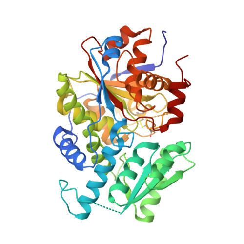
    Pyridoxal 5'-phosphate (PLP)-dependent enzymes utilize the unique chemistry of a pyridine ring to carry out diverse reactions involving amino acids. Diaminopropionate (DAP) ammonia-lyase (DAPAL) is a prokaryotic PLP-dependent enzyme that catalyzes the degradation of d- and l-forms of DAP to pyruvate and ammonia. Here, we report the first crystal structure of DAPAL from Escherichia coli (EcDAPAL) in tetragonal and monoclinic forms at 2.0 and 2.2 Å resolutions, respectively. Structures of EcDAPAL soaked with substrates were also determined. EcDAPAL has a typical fold type II PLP-dependent enzyme topology consisting of a large and a small domain with the active site at the interface of the two domains. The enzyme is a homodimer with a unique biological interface not observed earlier. Structure of the enzyme in the tetragonal form had PLP bound at the active site, whereas the monoclinic structure was in the apo-form. Analysis of the apo and holo structures revealed that the region around the active site undergoes transition from a disordered to ordered state and assumes a conformation suitable for catalysis only upon PLP binding. A novel disulfide was found to occur near a channel that is likely to regulate entry of ligands to the active site. EcDAPAL soaked with dl-DAP revealed density at the active site appropriate for the reaction intermediate aminoacrylate, which is consistent with the observation that EcDAPAL has low activity under crystallization conditions. Based on the analysis of the structure and results of site-directed mutagenesis, a two-base mechanism of catalysis involving Asp(120) and Lys(77) is suggested.


  • Organizational Affiliation
    • Molecular Biophysics Unit, Indian Institute of Science, Bangalore 560 012, India.

Macromolecule Content 

  • Total Structure Weight: 87.38 kDa 
  • Atom Count: 5,710 
  • Modeled Residue Count: 761 
  • Deposited Residue Count: 796 
  • Unique protein chains: 1

Macromolecules

Find similar proteins by:|  3D Structure
Entity ID: 1
MoleculeChains  Sequence LengthOrganismDetailsImage
Diaminopropionate ammonia-lyase
A, B
398Escherichia coli K-12Mutation(s): 0 
Gene Names: b2871JW2839ygeX
EC: 4.3.1.15
UniProt
Find proteins for P66899 (Escherichia coli (strain K12))
Explore P66899 
Go to UniProtKB:  P66899
Entity Groups
Sequence Clusters30% Identity50% Identity70% Identity90% Identity95% Identity100% Identity
UniProt GroupP66899
Sequence Annotations
Expand
Reference Sequence

Small Molecules

Ligands 1 Unique
IDChains Name / Formula / InChI Key2D Diagram3D Interactions
0JO

Query on 0JO


Download:Ideal Coordinates CCD File
C [auth A],
D [auth B]
2-{[(E)-{3-hydroxy-2-methyl-5-[(phosphonooxy)methyl]pyridin-4-yl}methylidene]amino}prop-2-enoic acid
C11 H13 N2 O7 P
BHIGINKEEFZJGX-YIXHJXPBSA-N

Experimental Data & Validation

Experimental Data

  • Method: X-RAY DIFFRACTION
  • Resolution: 2.50 Å
  • R-Value Free:  0.267 (Depositor), 0.271 (DCC) 
  • R-Value Work:  0.218 (Depositor), 0.215 (DCC) 
  • R-Value Observed: 0.220 (Depositor) 
Space Group: P 43 21 2
Unit Cell:
Length ( Å )Angle ( ˚ )
a = 85.506α = 90
b = 85.506β = 90
c = 205.084γ = 90
Software Package:
Software NamePurpose
MAR345dtbdata collection
PHASERphasing
REFMACrefinement
HKL-2000data reduction
SCALEPACKdata scaling

Structure Validation

View Full Validation Report


Ligand Structure Quality Assessment 


Entry History 

Deposition Data

Revision History  (Full details and data files)

  • Version 1.0: 2012-04-25
    Type: Initial release
  • Version 1.1: 2012-07-11
    Changes: Database references
  • Version 1.2: 2023-11-08
    Changes: Data collection, Database references, Derived calculations, Refinement description
  • Version 1.3: 2024-11-20
    Changes: Structure summary