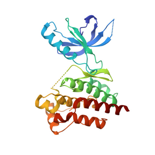
Allosteric Regulation of Focal Adhesion Kinase by Pip2 and ATP.
Zhou, J., Bronowska, A., Le Coq, J., Lietha, D., Grater, F.(2015) Biophys J 108: 698
- PubMed: 25650936
- DOI: https://doi.org/10.1016/j.bpj.2014.11.3454
- Primary Citation of Related Structures:
4D4R, 4D4S, 4D4V, 4D4Y, 4D55, 4D58, 4D5H, 4D5K - PubMed Abstract:
Focal adhesion kinase (FAK) is a nonreceptor tyrosine kinase that regulates cell signaling, proliferation, migration, and development. A major mechanism of regulation of FAK activity is an intramolecular autoinhibitory interaction between two of its domains--the catalytic and FERM domains. Upon cell adhesion to the extracellular matrix, FAK is being translocated toward focal adhesion sites and activated. Interactions of FAK with phosphoinositide phosphatidylinsositol-4,5-bis-phosphate (PIP₂) are required to activate FAK. However, the molecular mechanism of the activation remains poorly understood. Recent fluorescence resonance energy transfer experiments revealed a closure of the FERM-kinase interface upon ATP binding, which is reversed upon additional binding of PIP₂. Here, we addressed the allosteric regulation of FAK by performing all-atom molecular-dynamics simulations of a FAK fragment containing the catalytic and FERM domains, and comparing the dynamics in the absence or presence of ATP and PIP₂. As a major conformational change, we observe a closing and opening motion upon ATP and additional PIP₂ binding, respectively, in good agreement with the fluorescence resonance energy transfer experiments. To reveal how the binding of the regulatory PIP₂ to the FERM F2 lobe is transduced to the very distant F1/N-lobe interface, we employed force distribution analysis. We identified a network of mainly charged residue-residue interactions spanning from the PIP₂ binding site to the distant interface between the kinase and FERM domains, comprising candidate residues for mutagenesis to validate the predicted mechanism of FAK activation.
- Heidelberg Institute for Theoretical Studies, Heidelberg, Germany.
Organizational Affiliation:


