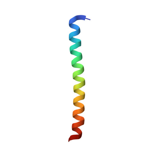
4D2H

Crystal structure of the tetramerisation domain of human CtIP


Experimental Data Snapshot

  • Method: X-RAY DIFFRACTION
  • Resolution: 1.90 Å
  • R-Value Free: 0.251 
  • R-Value Work: 0.212 
  • R-Value Observed: 0.214 

wwPDB Validation   3D Report Full Report


This is version 2.0 of the entry. See complete history


Literature

Ctip Tetramer Assembly is Required for DNA-End Resection and Repair.

Davies, O.R.Forment, J.V.Sun, M.Belotserkovskaya, R.Coates, J.Galanty, Y.Demir, M.Morton, C.R.Rzechorzek, N.J.Jackson, S.P.Pellegrini, L.

(2015) Nat Struct Mol Biol 22: 150

  • DOI: https://doi.org/10.1038/nsmb.2937
  • Primary Citation of Related Structures:  
    4D2H

  • PubMed Abstract: 

    Mammalian CtIP protein has major roles in DNA double-strand break (DSB) repair. Although it is well established that CtIP promotes DNA-end resection in preparation for homology-dependent DSB repair, the molecular basis for this function has remained unknown. Here we show by biophysical and X-ray crystallographic analyses that the N-terminal domain of human CtIP exists as a stable homotetramer. Tetramerization results from interlocking interactions between the N-terminal extensions of CtIP's coiled-coil region, which lead to a 'dimer-of-dimers' architecture. Through interrogation of the CtIP structure, we identify a point mutation that abolishes tetramerization of the N-terminal domain while preserving dimerization in vitro. Notably, we establish that this mutation abrogates CtIP oligomer assembly in cells, thus leading to strong defects in DNA-end resection and gene conversion. These findings indicate that the CtIP tetramer architecture described here is essential for effective DSB repair by homologous recombination.


  • Organizational Affiliation

    Department of Biochemistry, University of Cambridge, Cambridge, UK.


Macromolecules
Find similar proteins by:  (by identity cutoff)  |  3D Structure
Entity ID: 1
MoleculeChains Sequence LengthOrganismDetailsImage
RBBP8
A, B, C, D, E
A, B, C, D, E, F, G, H
38Homo sapiensMutation(s): 0 
EC: 3.1
UniProt & NIH Common Fund Data Resources
Find proteins for Q99708 (Homo sapiens)
Explore Q99708 
Go to UniProtKB:  Q99708
PHAROS:  Q99708
GTEx:  ENSG00000101773 
Entity Groups  
Sequence Clusters30% Identity50% Identity70% Identity90% Identity95% Identity100% Identity
UniProt GroupQ99708
Sequence Annotations
Expand
  • Reference Sequence
Small Molecules
Ligands 1 Unique
IDChains Name / Formula / InChI Key2D Diagram3D Interactions
SO4
Query on SO4

Download Ideal Coordinates CCD File 
I [auth A]
J [auth B]
K [auth B]
L [auth B]
M [auth C]
I [auth A],
J [auth B],
K [auth B],
L [auth B],
M [auth C],
N [auth D],
O [auth E],
P [auth F],
Q [auth F],
R [auth F],
S [auth G],
T [auth G],
U [auth G],
V [auth H]
SULFATE ION
O4 S
QAOWNCQODCNURD-UHFFFAOYSA-L
Experimental Data & Validation

Experimental Data

Unit Cell:
Length ( Å )Angle ( ˚ )
a = 35.97α = 90
b = 37.91β = 90.02
c = 97.79γ = 90
Software Package:
Software NamePurpose
BUSTERrefinement
XDSdata reduction
XDSdata scaling
Aimlessdata scaling
PHASERphasing

Structure Validation

View Full Validation Report


Entry History 

Deposition Data

Revision History  (Full details and data files)

  • Version 1.0: 2015-01-14
    Type: Initial release
  • Version 1.1: 2015-02-18
    Changes: Database references
  • Version 2.0: 2019-10-23
    Changes: Atomic model, Data collection, Other