4D05 | pdb_00004d05

Structure and activity of a minimal-type ATP-dependent DNA ligase from a psychrotolerant bacterium


Experimental Data Snapshot

  • Method: X-RAY DIFFRACTION
  • Resolution: 1.65 Å
  • R-Value Free: 
    0.192 (Depositor), 0.194 (DCC) 
  • R-Value Work: 
    0.145 (Depositor), 0.148 (DCC) 
  • R-Value Observed: 
    0.147 (Depositor) 

wwPDB Validation 3D Report Full Report

Validation slider image for 4D05

Ligand Structure Quality Assessment 


This is version 2.2 of the entry. See complete history

Literature

Enzyme-Adenylate Structure of a Bacterial ATP-Dependent DNA Ligase with a Minimized DNA-Binding Surface

Williamson, A.Rothweiler, U.Leiros, H.-K.S.

(2014) Acta Crystallogr D Biol Crystallogr 70: 3043

  • DOI: https://doi.org/10.1107/S1399004714021099
  • Primary Citation Related Structures: 
    4D05

  • PubMed Abstract: 

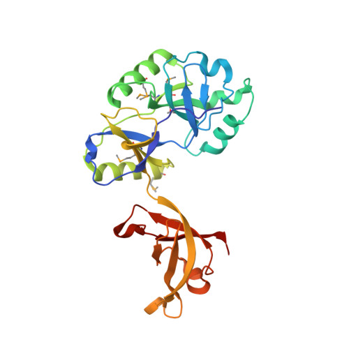
    DNA ligases are a structurally diverse class of enzymes which share a common catalytic core and seal breaks in the phosphodiester backbone of double-stranded DNA via an adenylated intermediate. Here, the structure and activity of a recombinantly produced ATP-dependent DNA ligase from the bacterium Psychromonas sp. strain SP041 is described. This minimal-type ligase, like its close homologues, is able to ligate singly nicked double-stranded DNA with high efficiency and to join cohesive-ended and blunt-ended substrates to a more limited extent. The 1.65 Å resolution crystal structure of the enzyme-adenylate complex reveals no unstructured loops or segments, and suggests that this enzyme binds the DNA without requiring full encirclement of the DNA duplex. This is in contrast to previously characterized minimal DNA ligases from viruses, which use flexible loop regions for DNA interaction. The Psychromonas sp. enzyme is the first structure available for the minimal type of bacterial DNA ligases and is the smallest DNA ligase to be crystallized to date.


  • Organizational Affiliation
    • Department of Chemistry, UiT The Arctic University of Norway, N-9037 Tromsø, Norway.

Macromolecule Content 

  • Total Structure Weight: 61.19 kDa 
  • Atom Count: 4,909 
  • Modeled Residue Count: 507 
  • Deposited Residue Count: 516 
  • Unique protein chains: 1

Macromolecules

Find similar proteins by:|  3D Structure
Entity ID: 1
MoleculeChains  Sequence LengthOrganismDetailsImage
ATP-DEPENDENT DNA LIGASE
A, B
258Psychromonas sp. SP041Mutation(s): 0 
UniProt
Find proteins for A0ACD6BAT9 (Psychromonas sp. SP041)
Explore A0ACD6BAT9 
Go to UniProtKB:  A0ACD6BAT9
Entity Groups
Sequence Clusters30% Identity50% Identity70% Identity90% Identity95% Identity100% Identity
UniProt GroupA0ACD6BAT9
Sequence Annotations
Expand
Reference Sequence

Small Molecules

Ligands 3 Unique
IDChains Name / Formula / InChI Key2D Diagram3D Interactions
AMP

Query on AMP


Download:Ideal Coordinates CCD File
H [auth A],
M [auth B]
ADENOSINE MONOPHOSPHATE
C10 H14 N5 O7 P
UDMBCSSLTHHNCD-KQYNXXCUSA-N
SO4

Query on SO4


Download:Ideal Coordinates CCD File
C [auth A]
D [auth A]
E [auth A]
F [auth A]
G [auth A]
C [auth A],
D [auth A],
E [auth A],
F [auth A],
G [auth A],
I [auth B],
J [auth B],
K [auth B],
L [auth B]
SULFATE ION
O4 S
QAOWNCQODCNURD-UHFFFAOYSA-L
MG

Query on MG


Download:Ideal Coordinates CCD File
N [auth B]MAGNESIUM ION
Mg
JLVVSXFLKOJNIY-UHFFFAOYSA-N
Modified Residues  1 Unique
IDChains TypeFormula2D DiagramParent
MSE
Query on MSE
A, B
L-PEPTIDE LINKINGC5 H11 N O2 SeMET

Experimental Data & Validation

Experimental Data

  • Method: X-RAY DIFFRACTION
  • Resolution: 1.65 Å
  • R-Value Free:  0.192 (Depositor), 0.194 (DCC) 
  • R-Value Work:  0.145 (Depositor), 0.148 (DCC) 
  • R-Value Observed: 0.147 (Depositor) 
Space Group: C 1 2 1
Unit Cell:
Length ( Å )Angle ( ˚ )
a = 178.427α = 90
b = 43.976β = 105.92
c = 89.633γ = 90
Software Package:
Software NamePurpose
PHENIXrefinement
XDSdata reduction
SHELXphasing

Structure Validation

View Full Validation Report


Ligand Structure Quality Assessment 


Entry History 

Deposition Data

Revision History  (Full details and data files)

  • Version 1.0: 2014-11-12
    Type: Initial release
  • Version 1.1: 2014-11-19
    Changes: Database references
  • Version 2.0: 2017-06-28
    Changes: Atomic model, Refinement description
  • Version 2.1: 2019-06-26
    Changes: Data collection, Database references, Derived calculations, Experimental preparation, Structure summary
  • Version 2.2: 2024-11-20
    Changes: Advisory, Data collection, Database references, Derived calculations, Other, Structure summary