4CWD | pdb_00004cwd

CRYSTAL STRUCTURE OF HUMAN GAMMA-BUTYROBETAINE,2-OXOGLUTARATE IN COMPLEX WITH 449, A NOVEL SUBSTRATE


Experimental Data Snapshot

  • Method: X-RAY DIFFRACTION
  • Resolution: 1.90 Å
  • R-Value Free: 
    0.206 (Depositor), 0.210 (DCC) 
  • R-Value Work: 
    0.167 (Depositor), 0.176 (DCC) 
  • R-Value Observed: 
    0.170 (Depositor) 

Starting Model: experimental
View more details

wwPDB Validation 3D Report Full Report

Validation slider image for 4CWD

Ligand Structure Quality Assessment 


This is version 1.4 of the entry. See complete history

Literature

Oxygenase-catalyzed desymmetrization of N,N-dialkyl-piperidine-4-carboxylic acids.

Rydzik, A.M.Leung, I.K.Kochan, G.T.McDonough, M.A.Claridge, T.D.Schofield, C.J.

(2014) Angew Chem Int Ed Engl 53: 10925-10927

  • DOI: https://doi.org/10.1002/anie.201406125
  • Primary Citation Related Structures: 
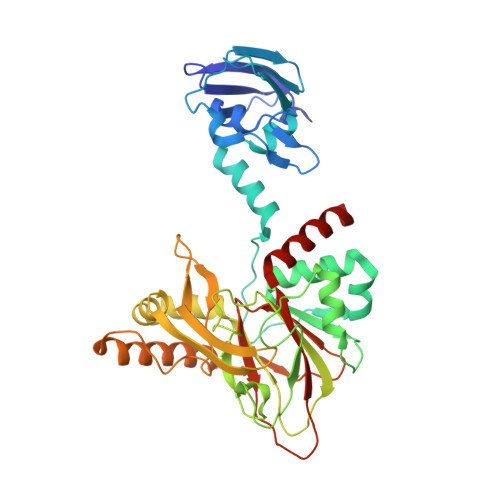
    4CWD

  • PubMed Abstract: 

    γ-Butyrobetaine hydroxylase (BBOX) is a 2-oxoglutarate dependent oxygenase that catalyzes the final hydroxylation step in the biosynthesis of carnitine. BBOX was shown to catalyze the oxidative desymmetrization of achiral N,N-dialkyl piperidine-4-carboxylates to give products with two or three stereogenic centers.


  • Organizational Affiliation
    • Department of Chemistry, University of Oxford, Chemistry Research Laboratory, 12 Mansfield Road, Oxford OX1 3TA (UK).

Macromolecule Content 

  • Total Structure Weight: 45.4 kDa 
  • Atom Count: 3,553 
  • Modeled Residue Count: 385 
  • Deposited Residue Count: 388 
  • Unique protein chains: 1

Macromolecules

Find similar proteins by:|  3D Structure
Entity ID: 1
MoleculeChains  Sequence LengthOrganismDetailsImage
GAMMA-BUTYROBETAINE DIOXYGENASE388Homo sapiensMutation(s): 0 
EC: 1.14.11.1
UniProt & NIH Common Fund Data Resources
Find proteins for O75936 (Homo sapiens)
Explore O75936 
Go to UniProtKB:  O75936
PHAROS:  O75936
GTEx:  ENSG00000129151 
Entity Groups
Sequence Clusters30% Identity50% Identity70% Identity90% Identity95% Identity100% Identity
UniProt GroupO75936
Sequence Annotations
Expand
Reference Sequence

Small Molecules

Ligands 5 Unique
IDChains Name / Formula / InChI Key2D Diagram3D Interactions
449

Query on 449


Download:Ideal Coordinates CCD File
D [auth A]4-carboxy-1,1-dimethylpiperidin-1-ium
C8 H16 N O2
ZUZDUOOCVXTRAU-UHFFFAOYSA-O
OGA

Query on OGA


Download:Ideal Coordinates CCD File
E [auth A]N-OXALYLGLYCINE
C4 H5 N O5
BIMZLRFONYSTPT-UHFFFAOYSA-N
PEG

Query on PEG


Download:Ideal Coordinates CCD File
F [auth A]DI(HYDROXYETHYL)ETHER
C4 H10 O3
MTHSVFCYNBDYFN-UHFFFAOYSA-N
ZN

Query on ZN


Download:Ideal Coordinates CCD File
C [auth A]ZINC ION
Zn
PTFCDOFLOPIGGS-UHFFFAOYSA-N
NI

Query on NI


Download:Ideal Coordinates CCD File
B [auth A]NICKEL (II) ION
Ni
VEQPNABPJHWNSG-UHFFFAOYSA-N

Experimental Data & Validation

Experimental Data

  • Method: X-RAY DIFFRACTION
  • Resolution: 1.90 Å
  • R-Value Free:  0.206 (Depositor), 0.210 (DCC) 
  • R-Value Work:  0.167 (Depositor), 0.176 (DCC) 
  • R-Value Observed: 0.170 (Depositor) 
Space Group: H 3 2
Unit Cell:
Length ( Å )Angle ( ˚ )
a = 106.518α = 90
b = 106.518β = 90
c = 205.347γ = 120
Software Package:
Software NamePurpose
PHENIXrefinement
DENZOdata reduction
SCALEPACKdata scaling
PHASERphasing

Structure Validation

View Full Validation Report


Ligand Structure Quality Assessment 


Entry History 

Deposition Data

Revision History  (Full details and data files)

  • Version 1.0: 2014-09-17
    Type: Initial release
  • Version 1.1: 2014-10-08
    Changes: Database references
  • Version 1.2: 2017-06-28
    Changes: Data collection
  • Version 1.3: 2018-03-07
    Changes: Database references, Source and taxonomy, Structure summary
  • Version 1.4: 2023-12-20
    Changes: Data collection, Database references, Derived calculations, Other, Refinement description